Native dataset
Data DOI: 10.15785/SBGRID/1241 | PDB ID 9Z7O: RCSB PDBe | Published: 26 May 2026
Corbett Laboratory, University of California, San Diego
Native dataset
Data DOI: 10.15785/SBGRID/1240 | PDB ID 9Z73: RCSB PDBe | Published: 26 May 2026
Corbett Laboratory, University of California, San Diego
Native dataset
Data DOI: 10.15785/SBGRID/1239 | PDB ID 9Z72: RCSB PDBe | Published: 26 May 2026
Corbett Laboratory, University of California, San Diego
Native dataset
Data DOI: 10.15785/SBGRID/1238 | PDB ID 9Z71: RCSB PDBe | Published: 26 May 2026
Corbett Laboratory, University of California, San Diego
Zinc anomalous SAD dataset
Data DOI: 10.15785/SBGRID/1156 | PDB ID 9MYP: RCSB PDBe | Published: 18 Aug 2025
Corbett Laboratory, University of California, San Diego
Zinc SAD dataset
Data DOI: 10.15785/SBGRID/1153 | PDB ID 9MUG: RCSB PDBe | Published: 18 Aug 2025
Corbett Laboratory, University of California, San Diego
native dataset
Data DOI: 10.15785/SBGRID/1145 | PDB ID 9EAV: RCSB PDBe | Published: 12 Sep 2025
Corbett Laboratory, University of California, San Diego
native dataset
Data DOI: 10.15785/SBGRID/1144 | PDB ID 9EAE: RCSB PDBe | Published: 12 Sep 2025
Corbett Laboratory, University of California, San Diego
native dataset
Data DOI: 10.15785/SBGRID/1143 | PDB ID 9EA8: RCSB PDBe | Published: 12 Sep 2025
Corbett Laboratory, University of California, San Diego
Native dataset
Data DOI: 10.15785/SBGRID/1142 | PDB ID 9EA5: RCSB PDBe | Published: 12 Sep 2025
Corbett Laboratory, University of California, San Diego
Native dataset
Data DOI: 10.15785/SBGRID/1141 | PDB ID 9EA4: RCSB PDBe | Published: 12 Sep 2025
Corbett Laboratory, University of California, San Diego
Single-wavelength dataset
Data DOI: 10.15785/SBGRID/1042 | PDB ID 8TYZ: RCSB PDBe | Published: 9 Jul 2024
Corbett Laboratory, University of California, San Diego
Single-wavelength dataset
Data DOI: 10.15785/SBGRID/1041 | PDB ID 8TZ0: RCSB PDBe | Published: 9 Jul 2024
Corbett Laboratory, University of California, San Diego
Single-wavelength dataset
Data DOI: 10.15785/SBGRID/1040 | PDB ID 8TYY: RCSB PDBe | Published: 9 Jul 2024
Corbett Laboratory, University of California, San Diego
Single-wavelength dataset
Data DOI: 10.15785/SBGRID/1039 | PDB ID 8TYX: RCSB PDBe | Published: 9 Jul 2024
Corbett Laboratory, University of California, San Diego
Native dataset to 1.77A
Data DOI: 10.15785/SBGRID/878 | PDB ID 7TSX: RCSB PDBe | Published: 6 Jan 2023
Corbett Laboratory, University of California, San Diego
Native dataset to 2.11 A resolution
Data DOI: 10.15785/SBGRID/877 | PDB ID 7TSQ: RCSB PDBe | Published: 6 Jan 2023
Corbett Laboratory, University of California, San Diego
Native data
Data DOI: 10.15785/SBGRID/868 | PDB ID 7T5V: RCSB PDBe | Published: 9 Sep 2022
Corbett Laboratory, University of California, San Diego
Native data
Data DOI: 10.15785/SBGRID/867 | PDB ID 7T5W: RCSB PDBe | Published: 9 Sep 2022
Corbett Laboratory, University of California, San Diego
Native data
Data DOI: 10.15785/SBGRID/866 | PDB ID 7T5U: RCSB PDBe | Published: 9 Sep 2022
Corbett Laboratory, University of California, San Diego
Selenomethione SAD (peak) data
Data DOI: 10.15785/SBGRID/865 | PDB ID 7T5T: RCSB PDBe | Published: 9 Sep 2022
Corbett Laboratory, University of California, San Diego
Native data
Data DOI: 10.15785/SBGRID/864 | PDB ID 7T5T: RCSB PDBe | Published: 9 Sep 2022
Corbett Laboratory, University of California, San Diego
Native dataset
Data DOI: 10.15785/SBGRID/837 | PDB ID 7R98: RCSB PDBe | Publication DOI: 10.3389/fimmu.2021.719037 | Published: 8 Feb 2022
Corbett Laboratory, University of California, San Diego
Process to 1.42 A
Data DOI: 10.15785/SBGRID/836 | PDB ID 7N0R: RCSB PDBe | Publication DOI: 10.3389/fimmu.2021.719037 | Published: 8 Feb 2022
Corbett Laboratory, University of California, San Diego
Native dataset
Data DOI: 10.15785/SBGRID/835 | PDB ID 7N0I: RCSB PDBe | Publication DOI: 10.3389/fimmu.2021.719037 | Published: 8 Feb 2022
Corbett Laboratory, University of California, San Diego
Zinc anomalous SAD dataset for V. polyspora Hop1 PHD-WHD domain
Data DOI: 10.15785/SBGRID/826 | PDB ID 7M0P: RCSB PDBe | Published: 4 Apr 2023
Corbett Laboratory, University of California, San Diego
Native dataset for SARS-CoV-2 Nucleocapsid (N) dimerization domain, P21 form
Data DOI: 10.15785/SBGRID/786 | PDB ID 6WZQ: RCSB PDBe | Published: 15 May 2020
Corbett Laboratory, University of California, San Diego
Native dataset for SARS-CoV-2 Nucleocapsid (N) dimerization domain, P1 form
Data DOI: 10.15785/SBGRID/785 | PDB ID 6WZO: RCSB PDBe | Published: 15 May 2020
Corbett Laboratory, University of California, San Diego
Native dataset for apo trimer form of Vibrio metoecus NucC
Data DOI: 10.15785/SBGRID/734 | PDB ID 6UXF: RCSB PDBe | Published: 20 Dec 2019
Corbett Laboratory, University of California, San Diego
Process to 1.45 A in XDS
Data DOI: 10.15785/SBGRID/732 | PDB ID 6Q1H: RCSB PDBe | Published: 20 Dec 2019
Corbett Laboratory, University of California, San Diego
Native dataset
Data DOI: 10.15785/SBGRID/711 | PDB ID 6U7B: RCSB PDBe | Published: 20 Dec 2019
Corbett Laboratory, University of California, San Diego
SAD dataset with selenomethionine-derivatized protein. One chain in AU; P6 symmetry reconstructs homo-hexamer
Data DOI: 10.15785/SBGRID/681 | PDB ID 6PB3: RCSB PDBe | Published: 20 Dec 2019
Corbett Laboratory, University of California, San Diego
Collected at Selenium peak wavelength with selenomethionine-derivatized protein, but no signal. Can process as a native dataset.
Data DOI: 10.15785/SBGRID/680 | PDB ID 6P8V: RCSB PDBe | Published: 20 Dec 2019
Corbett Laboratory, University of California, San Diego
Native dataset
Data DOI: 10.15785/SBGRID/679 | PDB ID 6P8U: RCSB PDBe | Published: 20 Dec 2019
Corbett Laboratory, University of California, San Diego
Native dataset
Data DOI: 10.15785/SBGRID/678 | PDB ID 6P8S: RCSB PDBe | Published: 20 Dec 2019
Corbett Laboratory, University of California, San Diego
Selenomethionine-derivatized SAD dataset
Data DOI: 10.15785/SBGRID/677 | PDB ID 6P8R: RCSB PDBe | Published: 20 Dec 2019
Corbett Laboratory, University of California, San Diego
Native dataset
Data DOI: 10.15785/SBGRID/676 | PDB ID 6P8R: RCSB PDBe | Published: 20 Dec 2019
Corbett Laboratory, University of California, San Diego
NaBr soak dataset (SAD)
Data DOI: 10.15785/SBGRID/674 | PDB ID 6P8P: RCSB PDBe | Published: 20 Dec 2019
Corbett Laboratory, University of California, San Diego
Native dataset
Data DOI: 10.15785/SBGRID/673 | PDB ID 6P8P: RCSB PDBe | Published: 20 Dec 2019
Corbett Laboratory, University of California, San Diego
Native dataset
Data DOI: 10.15785/SBGRID/672 | PDB ID 6P8O: RCSB PDBe | Published: 20 Dec 2019
Corbett Laboratory, University of California, San Diego
Native dataset
Data DOI: 10.15785/SBGRID/671 | PDB ID 6P8J: RCSB PDBe | Published: 20 Dec 2019
Corbett Laboratory, University of California, San Diego
Single-wavelength anomalous dataset (selenomethionine)
Data DOI: 10.15785/SBGRID/670 | PDB ID 6P82: RCSB PDBe | Published: 20 Dec 2019
Corbett Laboratory, University of California, San Diego
Single-wavelength selenomethionine anomalous dataset
Data DOI: 10.15785/SBGRID/669 | PDB ID 6P80: RCSB PDBe | Published: 20 Dec 2019
Corbett Laboratory, University of California, San Diego
Native dataset
Data DOI: 10.15785/SBGRID/668 | PDB ID 6P80: RCSB PDBe | Published: 20 Dec 2019
Corbett Laboratory, University of California, San Diego
450 frames, 0.2 degrees per frame
Data DOI: 10.15785/SBGRID/667 | PDB ID 6P7Q: RCSB PDBe | Published: 20 Dec 2019
Corbett Laboratory, University of California, San Diego
900 frames, 0.2 degrees per frame
Data DOI: 10.15785/SBGRID/666 | PDB ID 6P7P: RCSB PDBe | Published: 20 Dec 2019
Corbett Laboratory, University of California, San Diego
SAD data, two 30-degree wedges 180 degrees apart
Data DOI: 10.15785/SBGRID/665 | PDB ID 6P7O: RCSB PDBe | Published: 20 Dec 2019
Corbett Laboratory, University of California, San Diego
Native diffraction data
Data DOI: 10.15785/SBGRID/664 | PDB ID 6P7O: RCSB PDBe | Published: 20 Dec 2019
Corbett Laboratory, University of California, San Diego
Native dataset for C. glabrata Csm1:S. cerevisiae Dsn1(71-110) complex
Data DOI: 10.15785/SBGRID/610 | PDB ID 6MJE: RCSB PDBe | Published: 30 Oct 2018
Corbett Laboratory, University of California, San Diego
Native dataset for Candida glabrata Csm1:Dsn(43-67DD) complex
Data DOI: 10.15785/SBGRID/609 | PDB ID 6MJC: RCSB PDBe | Published: 30 Oct 2018
Corbett Laboratory, University of California, San Diego
Native dataset for Candida glabrata Csm1:Dsn1(14-72) complex
Data DOI: 10.15785/SBGRID/608 | PDB ID 6MJB: RCSB PDBe | Published: 30 Oct 2018
Corbett Laboratory, University of California, San Diego
Two native datasets from different crystals, which were indexed separately then scaled together to give the final dataset for refinement.
Data DOI: 10.15785/SBGRID/607 | PDB ID 6MJ8: RCSB PDBe | Published: 30 Oct 2018
Corbett Laboratory, University of California, San Diego
Zinc-anomalous SAD dataset for Eremothecium gossypii Shu1:Shu2 complex
Data DOI: 10.15785/SBGRID/586 | PDB ID 6DEX: RCSB PDBe | Published: 15 May 2018
Corbett Laboratory, University of California, San Diego
Single-wavelength dataset for S. cerevisiae Csm1-Dse3 complex
Data DOI: 10.15785/SBGRID/585 | PDB ID 6DEI: RCSB PDBe | Published: 25 Sep 2018
Corbett Laboratory, University of California, San Diego
Selenomethionine SAD dataset for M. musculus SYCP3 105-248, P1 crystal form
Data DOI: 10.15785/SBGRID/584 | PDB ID 6DD9: RCSB PDBe | Published: 25 Jan 2019
Corbett Laboratory, University of California, San Diego
Selenomethionine SAD dataset for M. musculus SYCP3 105-248, P21 crystal form
Data DOI: 10.15785/SBGRID/583 | PDB ID 6DD8: RCSB PDBe | Published: 25 Jan 2019
Corbett Laboratory, University of California, San Diego
SeMet SAD dataset, C2 crystal form
Data DOI: 10.15785/SBGRID/540 | PDB ID 6BZG: RCSB PDBe | Published: 6 Feb 2018
Corbett Laboratory, University of California, San Diego
Native dataset, P212121 crystal form
Data DOI: 10.15785/SBGRID/539 | PDB ID 6BZG: RCSB PDBe | Published: 6 Feb 2018
Corbett Laboratory, University of California, San Diego
Native dataset, C2 crystal form
Data DOI: 10.15785/SBGRID/538 | PDB ID 6BZF: RCSB PDBe | Published: 6 Feb 2018
Corbett Laboratory, University of California, San Diego
Native dataset for H. sapiens TRIP13 E253Q + ATP
Data DOI: 10.15785/SBGRID/410 | PDB ID 5VQA: RCSB PDBe | Published: 6 Jun 2017
Corbett Laboratory, University of California, San Diego
Native dataset for hTRIP13 E253Q Apo state
Data DOI: 10.15785/SBGRID/409 | PDB ID 5VQ9: RCSB PDBe | Published: 6 Jun 2017
Corbett Laboratory, University of California, San Diego
Native dates for Csm1 bound to Ulp2-Tof2 fusion
Data DOI: 10.15785/SBGRID/398 | PDB ID 53VN: RCSB PDBe | Published: 9 May 2017
Corbett Laboratory, University of California, San Diego
Native data for PDB entry 5SZC
Data DOI: 10.15785/SBGRID/359 | PDB ID 5SZC: RCSB PDBe | Published: 5 Sep 2017
Corbett Laboratory, University of California, San Diego
Native data for PDB 5SZB
Data DOI: 10.15785/SBGRID/358 | PDB ID 5SZB: RCSB PDBe | Published: 5 Sep 2017
Corbett Laboratory, University of California, San Diego
Zinc SAD data for PDB entry 5SZB
Data DOI: 10.15785/SBGRID/357 | PDB ID 5SZB: RCSB PDBe | Published: 5 Sep 2017
Corbett Laboratory, University of California, San Diego
Native dataset for S. cerevisiae Csm1:Ulp2 complex
Data DOI: 10.15785/SBGRID/327 | PDB ID 5V1A: RCSB PDBe | Published: 9 May 2017
Corbett Laboratory, University of California, San Diego
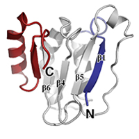
Native data for C. elegans HTP-2 bound to HIM-3 closure motif, P212121 form
Data DOI: 10.15785/SBGRID/210 | PDB ID 4TZS: RCSB PDBe | Publication DOI: 10.1016/j.devcel.2014.09.013 | Published: 18 Dec 2015
Corbett Laboratory, University of California, San Diego
Native data for C. elegans HTP-1 bound to HTP-3 motif-1
Data DOI: 10.15785/SBGRID/209 | PDB ID 4TZQ: RCSB PDBe | Publication DOI: 10.1016/j.devcel.2014.09.013 | Published: 18 Dec 2015
Corbett Laboratory, University of California, San Diego
Native data for C. elegans HTP-1 bound to HIM-3 closure motif
Data DOI: 10.15785/SBGRID/208 | PDB ID 4TZO: RCSB PDBe | Publication DOI: 10.1016/j.devcel.2014.09.013 | Published: 18 Dec 2015
Corbett Laboratory, University of California, San Diego
Native data for C. elegans HTP-2 bound to HTP-3 motif-6
Data DOI: 10.15785/SBGRID/207 | PDB ID 4TZN: RCSB PDBe | Publication DOI: 10.1016/j.devcel.2014.09.013 | Published: 18 Dec 2015
Corbett Laboratory, University of California, San Diego
Native data for C. elegans HTP-2 bound to HTP-3 closure motif 1
Data DOI: 10.15785/SBGRID/206 | PDB ID 4TZM: RCSB PDBe | Publication DOI: 10.1016/j.devcel.2014.09.013 | Published: 18 Dec 2015
Corbett Laboratory, University of California, San Diego
Native data for C. elegans HTP-2 bound to HIM-3 closure motif, P21 form
Data DOI: 10.15785/SBGRID/205 | PDB ID 4TZL: RCSB PDBe | Publication DOI: 10.1016/j.devcel.2014.09.013 | Published: 18 Dec 2015
Corbett Laboratory, University of California, San Diego
Native data for C. elegans HIM-3 bound to HTP-3 closure motif-4
Data DOI: 10.15785/SBGRID/204 | PDB ID 4TZJ: RCSB PDBe | Publication DOI: 10.1016/j.devcel.2014.09.013 | Published: 18 Dec 2015
Corbett Laboratory, University of California, San Diego
Native dataset for Hrr25:Mam1 complex, form 2
Data DOI: 10.15785/SBGRID/196 | PDB ID 5CZO: RCSB PDBe | Published: 9 Aug 2016
Corbett Laboratory, University of California, San Diego
Native diffraction data for S. cerevisiae Hrr25:Mam1, form 1
Data DOI: 10.15785/SBGRID/195 | PDB ID 5CYZ: RCSB PDBe | Published: 9 Aug 2016
Corbett Laboratory, University of California, San Diego
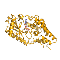
S. cerevisiae Hrr25 bound to inhibitor CK1-7
Data DOI: 10.15785/SBGRID/154 | PDB ID 4XHL: RCSB PDBe | Published: 9 Aug 2016
Corbett Laboratory, University of California, San Diego
C. glabrata Hrr25 in Apo state
Data DOI: 10.15785/SBGRID/153 | PDB ID 4XHH: RCSB PDBe | Published: 9 Aug 2016
Corbett Laboratory, University of California, San Diego
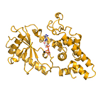
C. glabrata Hrr25 bound to ADP in Formate buffer
Data DOI: 10.15785/SBGRID/152 | PDB ID 4XHG: RCSB PDBe | Published: 9 Aug 2016
Corbett Laboratory, University of California, San Diego
C. glabrata Hrr25 bound to ADP in Ammonium sulfate buffer
Data DOI: 10.15785/SBGRID/151 | PDB ID 4XH0: RCSB PDBe | Published: 9 Aug 2016
Corbett Laboratory, University of California, San Diego
Selenomethionine dataset for C. elegans PCH-2
Data DOI: 10.15785/SBGRID/21 | PDB ID 4XGU: RCSB PDBe | Publication DOI: 10.7554/eLife.07367 | Published: 6 May 2015
Corbett Laboratory, University of California, San Diego
Native dataset of C. elegans PCH-2, crystallized without addition of nucleotides
Data DOI: 10.15785/SBGRID/20 | PDB ID 4XGU: RCSB PDBe | Publication DOI: 10.7554/eLife.07367 | Published: 6 May 2015
Corbett Laboratory, University of California, San Diego
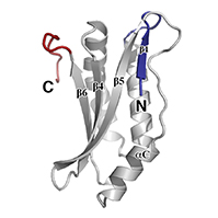
Selenomethionine SAD dataset for C. elegans HIM-3
Data DOI: 10.15785/SBGRID/17 | PDB ID 4TRK: RCSB PDBe | Publication DOI: 10.1016/j.devcel.2014.09.013 | Published: 5 May 2015
Corbett Laboratory, University of California, San Diego
C. elegans HIM-3 full-length crystallized in sodium malonate
Data DOI: 10.15785/SBGRID/16 | PDB ID 4TRK: RCSB PDBe | Publication DOI: 10.1016/j.devcel.2014.09.013 | Published: 5 May 2015
Corbett Laboratory, University of California, San Diego