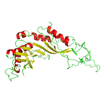
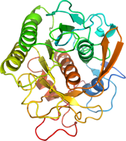
Continous Serial Electron Diffraction Data of MutT homolog 1 and its substrate 8-oxo-dGTP
Data DOI: 10.15785/SBGRID/1163 | Published: 6 Jan 2026
Zou Laboratory, Stockholm University
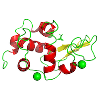
Serial electron diffraction (SerialED) data of Urate Oxidase from Aspergillus flavus complexed with uric acid
Data DOI: 10.15785/SBGRID/1157 | Published: 6 Jan 2026
Zou Laboratory, Stockholm University
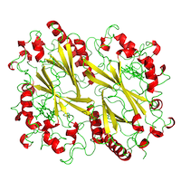
Serial electron diffraction (SerialED) data of Urate Oxidase from Aspergillus flavus complexed with 9-methyl uric acid
Data DOI: 10.15785/SBGRID/1154 | Published: 6 Jan 2026
Zou Laboratory, Stockholm University
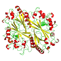
Serial electron diffraction (SerialED) data of triclinic hen egg-white lysozyme
Data DOI: 10.15785/SBGRID/1150 | Published: 6 Jan 2026
Zou Laboratory, Stockholm University
Serial electron diffraction (SerialED) data of Dye Type Peroxidase Aa from Streptomyces lividans
Data DOI: 10.15785/SBGRID/1149 | PDB ID 9FYK: RCSB PDBe | Published: 6 Jan 2026
Zou Laboratory, Stockholm University
Serial electron diffraction (SerialED) data of Dye Type Peroxidase Aa from Streptomyces lividans complexed with azide (N3-)
Data DOI: 10.15785/SBGRID/1146 | PDB ID 9FY7: RCSB PDBe | Published: 6 Jan 2026
Zou Laboratory, Stockholm University
(2S,3aS,8aS)-8a-hydroxy-3a-methyl-1,2,3,3a,8,8a-hexahydroindeno[2,1-b]pyrrole-2-carboxylic acid monohydrate
Data DOI: 10.15785/SBGRID/979 | Publication DOI: 10.1021/jacs.9b09864 | Published: 6 Jan 2023
Gonen Laboratory, University of California, Los Angeles
Ab initio structure of proteinase K from electron-counted MicroED data
Data DOI: 10.15785/SBGRID/978 | PDB ID 7SKX: RCSB PDBe | Publication DOI: 10.1038/s41592-022-01485-4 | Published: 3 Jan 2023
Gonen Laboratory, University of California, Los Angeles
MicroED structure of proteinase K from a platinum-coated, polished, single lamella at 1.79 Å resolution
Data DOI: 10.15785/SBGRID/977 | PDB ID 6PKR: RCSB PDBe | Publication DOI: 10.1016/j.str.2019.07.004 | Published: 3 Jan 2023
Gonen Laboratory, University of California, Los Angeles
MicroED data of tetragonal lysozyme collected from specimens prepared by Vitrobot and Preassis.
Data DOI: 10.15785/SBGRID/842 | Published: 9 Jul 2021
Zou Laboratory, Stockholm University
MicroED structure of a natural product VFAThiaGlu
Data DOI: 10.15785/SBGRID/820 | PDB ID 6PO6: RCSB PDBe | Publication DOI: 10.1126/science.aau6232 | Published: 29 Jan 2021
Gonen Laboratory, University of California, Los Angeles
1.01 Å MicroED structure of GSNQNNF at 0.27 e- / A^2
Data DOI: 10.15785/SBGRID/819 | PDB ID 6CLC: RCSB PDBe | Publication DOI: 10.1016/j.str.2018.03.021 | Published: 29 Jan 2021
Gonen Laboratory, University of California, Los Angeles
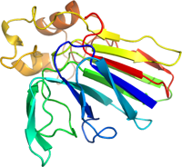
MicroED data of MyD88 TIR domain higher-order assembly microcrystals
Data DOI: 10.15785/SBGRID/814 | PDB ID 7BEQ: RCSB PDBe | Published: 12 Mar 2021
Zou Laboratory, Stockholm University
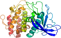
Microcrystal electron diffraction data of HCA II
Data DOI: 10.15785/SBGRID/793 | PDB ID 6YMB: RCSB PDBe | Published: 4 Aug 2020
Zou Laboratory, Stockholm University
Microcrystal electron diffraction data of HCA II:AZM
Data DOI: 10.15785/SBGRID/792 | PDB ID 6YMA: RCSB PDBe | Published: 4 Aug 2020
Zou Laboratory, Stockholm University
MicroED structure of a FIB-milled CypA Crystal; Tamir Gonen, Michael C Thompson, and James Fraser are co-PIs of this dataset.
Data DOI: 10.15785/SBGRID/752 | PDB ID 6U5G: RCSB PDBe | Published: 28 Jan 2020
Fraser Laboratory, University of California, San Francisco
Micro-electron diffraction data from thermolysin
Data DOI: 10.15785/SBGRID/290 | PDB ID 5k7t: RCSB PDBe | Published: 31 Mar 2017
Gonen Laboratory, University of California, Los Angeles
Micro-electron diffraction data from proteinase K
Data DOI: 10.15785/SBGRID/289 | PDB ID 5k7s: RCSB PDBe | Published: 31 Mar 2017
Gonen Laboratory, University of California, Los Angeles
Micro-electron diffraction data from trypsin
Data DOI: 10.15785/SBGRID/288 | PDB ID 5k7r: RCSB PDBe | Published: 31 Mar 2017
Gonen Laboratory, University of California, Los Angeles
Micro-electron diffraction data from thaumatin
Data DOI: 10.15785/SBGRID/287 | PDB ID 5k7q: RCSB PDBe | Published: 31 Mar 2017
Gonen Laboratory, University of California, Los Angeles