XDS processed intensities generously provided by Kay Diederichs and Tom Terwilliger.
Zn-SAD dataset
Data DOI: 10.15785/SBGRID/3 | PDB ID 3TRZ: RCSB PDBe | Publication DOI: 10.1016/j.cell.2011.10.020 | Published: 13 Apr 2015
Sliz Laboratory, Harvard Medical School
Native
Data DOI: 10.15785/SBGRID/5 | PDB ID 4TKN: RCSB PDBe | Publication DOI: 10.1074/jbc.M114.584011 | Published: 17 Apr 2015
Boggon Laboratory, Yale University School of Medicine
Collection temperature 100K Wavelength 1.1271Å
Data DOI: 10.15785/SBGRID/9 | PDB ID 4NWV: RCSB PDBe | Publication DOI: 10.1073/pnas.1407122111 | Published: 20 Apr 2015
Tao Laboratory, Rice University
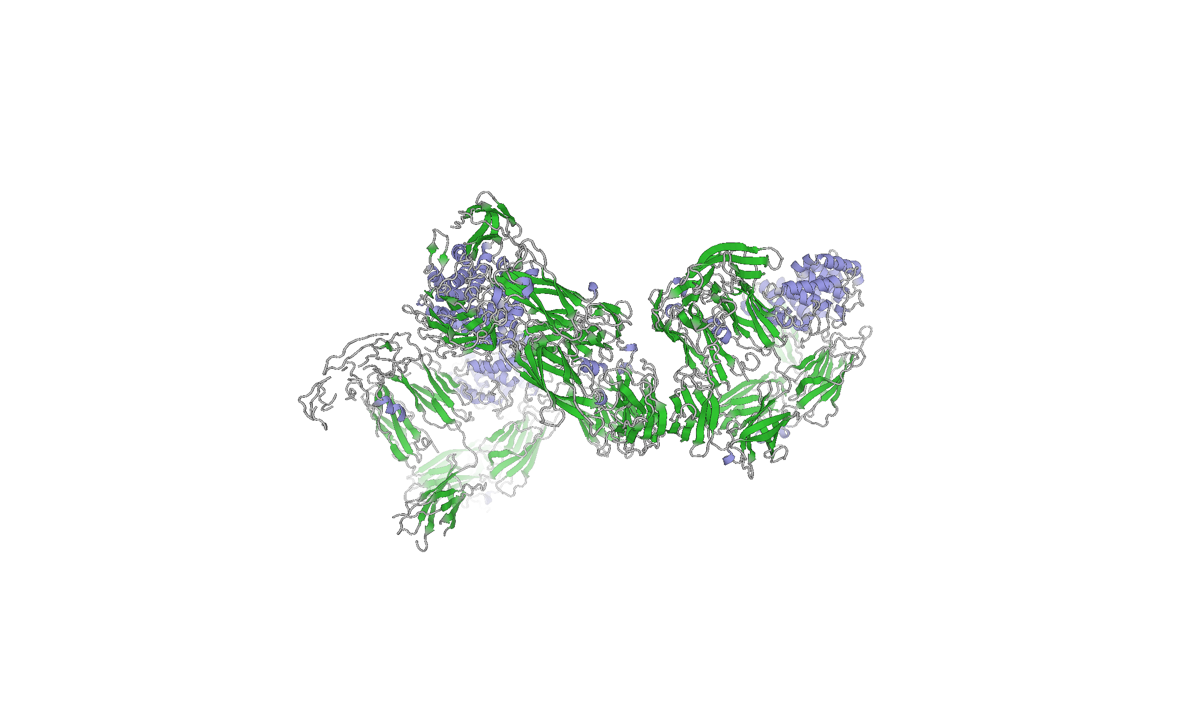
Uranyl acetate derivative 2, E2 domains I-II
Data DOI: 10.15785/SBGRID/62 | PDB ID 4ILD: RCSB PDBe | Publication DOI: 10.1073/pnas.1300524110 | Published: 7 May 2015
Modis Laboratory, University of Cambridge
Highest resolution room temperature diffraction data set for Cyclophilin A. Collected under paratone oil.
Data DOI: 10.15785/SBGRID/68 | PDB ID 4YUO: RCSB PDBe | Publication DOI: 10.1101/016733 | Published: 8 May 2015
Fraser Laboratory, University of California, San Francisco
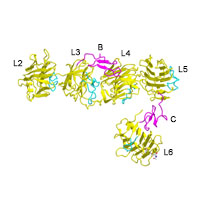
native high resolution data set used for final refinement of structure 3QCW; 5 wedges of 125 frames each, 0.5 degree oscillation angle; collected at 1.12718 Ang (10 999.5 eV); XD 300 mm MarMosaic CCD 300 detector; beam position (x: 150.0 mm , y: 150.0 mm)
Data DOI: 10.15785/SBGRID/78 | PDB ID 3QCW: RCSB PDBe | Published: 11 May 2015
Rudenko Laboratory, University of Texas Medical Branch
Native dataset
Data DOI: 10.15785/SBGRID/83 | PDB ID 4XC6: RCSB PDBe | Publication DOI: 10.1073/pnas.1419582112 | Published: 13 May 2015
Drennan Laboratory, Massachusetts Institute of Technology
This is a three wavelength MAD data collected at the iron edge. E1 = 7134.03 eV; E2 = 7634 eV; E3 = 7124.35 eV. Bijvoet pairs were collected in real time using a 1 deg wedge (inverse beam), which is the standard protocol at ALS beamline 8.3.1. Data identifier: Sono: but1_1_E1_### to but1_1_E3_### The crystal used here yields a complete 2.88 A MAD data set.
Data DOI: 10.15785/SBGRID/97 | PDB ID 1XBN: RCSB PDBe | Publication DOI: 10.1126/science.1103596 | Published: 19 May 2015
Raman Laboratory, University of Maryland
Diffraction dataset for 'Spinach' RNA-chromophore complex
Data DOI: 10.15785/SBGRID/111 | PDB ID 4TS0: RCSB PDBe | Publication DOI: 10.1038/nsmb.2865 | Published: 19 May 2015
Ferré-D'Amaré Laboratory, National Institutes of Health
Native dataset
Data DOI: 10.15785/SBGRID/117 | PDB ID 4LNV: RCSB PDBe | Publication DOI: 10.1371/journal.ppat.1002958 | Published: 5 Jun 2015
Baxter Laboratory, Yale University
This is a selenomethionine single-wavelength anomalous diffraction data set collected at 0.9789 angstrom wavelength
Data DOI: 10.15785/SBGRID/123 | PDB ID 3M1C: RCSB PDBe | Publication DOI: 10.1038/nsmb.1837 | Published: 21 May 2015
Heldwein Laboratory, Tufts University School of Medicine
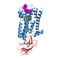
This dataset is compiled from 18 crystals of M2 receptor grown in the presence of the agonist iperoxo and the allosteric modulator LY2119620.
Data DOI: 10.15785/SBGRID/125 | PDB ID 4MQT: RCSB PDBe | Publication DOI: 10.1038/nature12735 | Published: 21 May 2015
Kruse Laboratory, Harvard Medical School
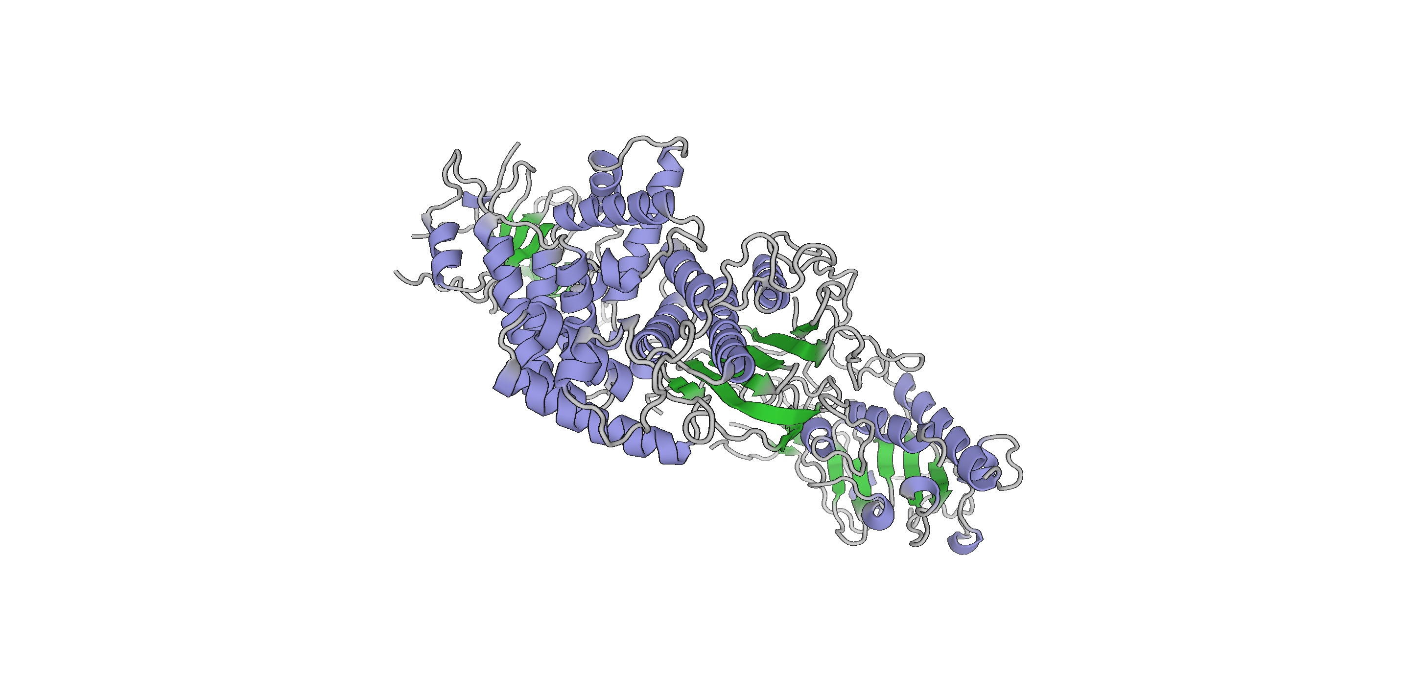
native data, collected in two separate spots
Data DOI: 10.15785/SBGRID/179 | PDB ID 4FHN: RCSB PDBe | Publication DOI: 10.1073/pnas.1205151109 | Published: 6 Oct 2015
Schwartz Laboratory, Massachusetts Institute of Technology
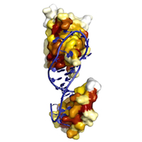
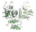
selenomethionyl neurexin 1alpha ectodomain
Data DOI: 10.15785/SBGRID/218 | PDB ID 3QCW: RCSB PDBe | Publication DOI: 10.1016/j.str.2011.03.012 | Published: 12 Jan 2016
Rudenko Laboratory, University of Texas Medical Branch