The set of all AlphaFold multimer (AF-M) v2.3 pairwise structure predictions accompanying the publication: Predictomes: A classifier-curated database of AlphaFold-modeled protein-protein interactions. This dataset includes prediction pairs used for training random forest classifiers including SPOC, pairs used for 30 ranking experiments, all pairs that belong to the genome maintenance matrix on predictomes.org, and three proteome wide in-silico interaction screens conducted with human DONSON, human STK19, and human USP37. All pairs were generated with ColabFold v1.5.2. All our predictions used AF-M multimer version 3 weights models 1, 2, and 4 with 3 recycles, templates enabled, 1 ensemble, no dropout, and no AMBER relaxation. The Multiple Sequence Alignments (MSAs) (unpaired + paired) supplied to AF-M were generated by the MMSeqs2 server using default settings. Sequences run were generally capped at 3,600 amino acids total to avoid memory exhaustion on GPUs.
Data DOI: 10.15785/SBGRID/1155 | Published: 4 Feb 2025
Walter Laboratory, Harvard Medical School
Antibody-antigen models for the paper "Towards the accurate modelling of antibody-antigen complexes from sequence using machine learning and information-driven docking". M Giulini, C Schneider, D Cutting, N Desai, C Deane, AMJJ Bonvin. Bioinformatics. 2024
Data DOI: 10.15785/SBGRID/1139 | Published: 16 Feb 2025
Bonvin Laboratory, Utrecht University
Protein-glycan docking models obtained with HADDOCK corresponding to the protocols described in: A Ranaudo, M Giulini, A Pelissou Ayuso, AMJJ Bonvin. Modelling Protein-Glycan Interactions with HADDOCK. Journal of Chemical Information and Modeling (2024)
Data DOI: 10.15785/SBGRID/1138 | Published: 12 Feb 2025
Bonvin Laboratory, Utrecht University
Protein-cyclic peptides docking models obtained with HADDOCK corresponding to the optimal protocol described in: V. Charitou, S.C. van Keulen and A.M.J.J. Bonvin. A Cyclisation and Docking Protocol for Cyclic Peptide-Protein Modelling using HADDOCK2.4. J. Chem. Theo. and Comp. (2022) - https://doi.org/10.1021/acs.jctc.2c00075
Data DOI: 10.15785/SBGRID/912 | Publication DOI: https://doi.org/10.1021/acs.jctc.2c00075 | Published: 7 May 2024
Bonvin Laboratory, Utrecht University
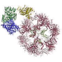
Structural model of the E3BP core, refined in a C2 symmetrized cryoEM map. E3BP showed a minimal fold, which is conserved, and can be found in various other E2 proteins from diverse acyl-transferases, including the 2-keto acid dehydrogenase family. Additionally, by fitting the structural models into an asymmetrically refined cryoEM map, we supply a structural model for the native core scaffold of the PDHc metabolon from C. thermophilum. Models were built and refined by COOT and PHENIX. To capture the transient interaction of lipoyl domain (LD) and the core structure during the transacetylase reaction, we docked the LDs of C. thermophilum, H. sapiens, and N. crassa to their respective core structure. HADDOCK parameter files are deposited to reproduce docking. The best docking solution of C. thermophilum was used to study the interaction by extensive MD simulations. Parameter files and results are also given, to reproduce these simulations.
Data DOI: 10.15785/SBGRID/848 | PDB ID 7OTT: RCSB PDBe | Published: 23 Nov 2021
Kastritis Laboratory, Martin Luther University Halle-Wittenberg
HADDOCK refined 3D structures from multiple datasets (BM5, MANY, DC, CAPRI), used for experiments in the DeepRank paper. 1.Renaud, N. et al. DeepRank: A deep learning framework for data mining 3D protein-protein interfaces. bioRxiv 2021.01.29.425727 (2021) doi:10.1101/2021.01.29.425727.
Data DOI: 10.15785/SBGRID/843 | Published: 28 Oct 2021
Xue Laboratory, Radboud University Medical Center
This dataset comprises bound TCR-pMHC models for 44 TCR docking benchmark cases. These models were produced using four docking platforms - ClusPro, HADDOCK, LightDock and ZDOCK - as a comparative study of docking software performance in the context of TCR-pMHC modelling. Each docking case was provided to the software platforms along with varying levels of detail about the binding interface in the form of four docking scenarios, to assess how effectively each platform made use of this additional information to improve modeling accuracy. A manuscript reporting these results has been submitted for publication.
Data DOI: 10.15785/SBGRID/825 | Published: 21 May 2021
Chain Laboratory, University College London
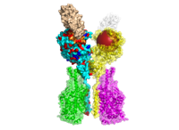
Structural models of the E1 and E2 proteins of Pyruvate Dehydrogenase Complex (PDHc), 2-Oxoglutarate Dehydrogenase (OGDHc), and Branched-Chain alpha-Keto Acid Dehydrogenase Complex (BCKDHc), E3BP of PDHc and E3, shared among all three complexes. In addition, a cif-file of E1, E2, E3BP, and E3 of PDHc modeled from cryoEM data is provided. Models were generated by homology modeling using MODELLER and refined using HADDOCK webserver.
Data DOI: 10.15785/SBGRID/817 | PDB ID 7BGJ: RCSB PDBe | Published: 26 Jan 2021
Kastritis Laboratory, Martin Luther University Halle-Wittenberg
242 structural models of human ACE2 variants with the bound RBD of SARS-CoV-2 Spike glycoprotein refined with HADDOCK2.2 (initial PDB: 6M17, 6M0J). The structural models include in human population naturally occurring variants of ACE2 (140), models of the mutants with reported effects on the recognition of RBD (39), and computational alanine scanning mutagenesis of ACE2-RBD interface (63). Moreover, all aforementioned ACE2 variants can be found as a structural models of ACE2-B(0)AT1 complex with the bound RBD, including all ions and structurally-important glycan molecules (initial PDB: 6M17).
Data DOI: 10.15785/SBGRID/791 | Published: 21 Aug 2020
Kastritis Laboratory, Martin Luther University Halle-Wittenberg
Simulated lysozyme data set - the diffraction images were generated by MLFSOM. The calculation of structure factors was based on the gadolinium derivative of tetragonal Hen Egg-White Lysozyme (PDB 1H87), all the alternative conformations of residues were removed.
Data DOI: 10.15785/SBGRID/746 | Published: 29 May 2020
Dohnalek Laboratory, Institute of Biotechnology of the Czech Academy of Sciences
PDBs, scores and RMSDs related with ClustENM-HADDOCK modelling pipeline for protein-protein and protein-DNA complexes
Data DOI: 10.15785/SBGRID/707 | Published: 27 Sep 2019
Bonvin Laboratory, Utrecht University
Antibody-antigen docking models gathered for the article: "Modelling of antibody-antigen complexes by information-driven docking." F. Ambrosetti, B. Jimenez-Garcia, J. Roel-Touris, A.M.J.J. Bonvin. Jun 2019. Structure.
Data DOI: 10.15785/SBGRID/686 | Published: 27 Sep 2019
Bonvin Laboratory, Utrecht University
HADDOCK docking models for Protein-Protein Docking Benchmark 4; HADDOCK, pyDock, SwarmDock and ZDock docking models for new complexes of Docking Benchmark 5; Various docking models for CAPRI score_set. All of the non-HADDOCK models are refined with HADDOCK using energy minimisation.
Data DOI: 10.15785/SBGRID/684 | Publication DOI: 10.1093/bioinformatics/btz496 | Published: 9 Jul 2019
Bonvin Laboratory, Utrecht University
HADDOCK models of mutant protein complexes gathered for the article: "C. Geng, A. Vangone, G.E. Folkers, L.C. Xue and A.M.J.J. Bonvin. iSEE: Interface Structure, Evolution and Energy-based machine learning predictor of binding affinity changes upon mutations. Proteins: Struc. Funct. & Bioinformatics 87, 110-119 (2019)."
Data DOI: 10.15785/SBGRID/651 | Publication DOI: 10.1002/prot.25630 | Published: 9 Jul 2019
Bonvin Laboratory, Utrecht University
Decoys of a membrane protein complex docking benchmark. The decoys were obtained after docking with the HADDOCK webserver (v2.2) and they belong in two sets which reflect two extreme docking scenarios. One where we would have no information about the nature of the interaction and we use random restraints to drive the docking, and one where we use restraints extracted from the interface of the native complex to drive the docking. We have generated 50800 structures for the first scenario, distributed in three stages: 50000, 400 and 400 for the rigid-body, simulated annealing and flexible refinement stages respectively. We have generated 10800 structures for the second scenario distributed in three stages: 10000, 400 and 400 for the rigid-body, simulated annealing and flexible refinement stages respectively.
Data DOI: 10.15785/SBGRID/618 | Published: 29 Oct 2018
Bonvin Laboratory, Utrecht University
HADDOCK refined models for the biological/crystallographic interfaces collected in the DC and MANY datasets
Data DOI: 10.15785/SBGRID/566 | Published: 6 Mar 2018
Bonvin Laboratory, Utrecht University
HADDOCK models of mutant protein complexes gathered for the article: "C. Geng, A. Vangone, G.E. Folkers, L. Xue and Alexandre M.J.J. Bonvin, iSEE: Interface Structure, Evolution and Energy-based random forest predictor of binding affinity changes upon mutations. 2017. Submitted".
Data DOI: 10.15785/SBGRID/520 | Published: 5 Dec 2017
Bonvin Laboratory, Utrecht University
HADDOCK protein-peptide models gathered for the article: "A unified conformational selection and induced fit approach to protein-peptide docking." Trellet M, Melquiond ASJ, Bonvin AMJJ. Mar 2013. PLoS One 8(3) dx.doi.org/10.1371/journal.pone.0058769
Data DOI: 10.15785/SBGRID/420 | Publication DOI: 10.1371/journal.pone.0058769 | Published: 24 Jan 2017
Bonvin Laboratory, Utrecht University
This is a correction of the folder of CAPRI30 in the original SBgrid 221 dataset
Data DOI: 10.15785/SBGRID/261 | Publication DOI: 10.1093/bib/bbw027 | Published: 26 Apr 2016
Bonvin Laboratory, Utrecht University
THIS DATA IS ORIGINALLY USED IN XUE ET AL., BRIEFINGS IN BIOINFORMATICS, 2016: TEMPLATE-BASED PROTEIN-PROTEIN DOCKING EXPLOITING PAIRWISE INTERFACIAL RESIDUE RESTRAINTS by Li C Xue, Joao P.G.L.M. Rodrigues, Drena Dobbs, Vasant Honavar, Alexandre M.J.J. Bonvin
Data DOI: 10.15785/SBGRID/221 | Publication DOI: 10.1093/bib/bbw027 | Published: 9 Mar 2016
Bonvin Laboratory, Utrecht University