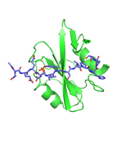
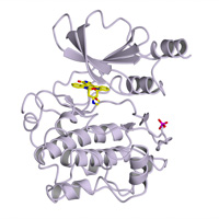
Two native datasets from one crystal were merged together by rescaling using XSCALE.
Data DOI: 10.15785/SBGRID/1187 | PDB ID 9P6A: RCSB PDBe | Publication DOI: 10.1038/s41467-025-66884-5 | Published: 14 Nov 2025
Boggon Laboratory, Yale University School of Medicine
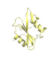
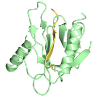
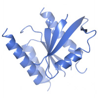
X-Ray Diffraction data for c-Src SH3 domain
Data DOI: 10.15785/SBGRID/1158 | PDB ID 9OFX: RCSB PDBe | Published: 2 May 2025
Boggon Laboratory, Yale University School of Medicine
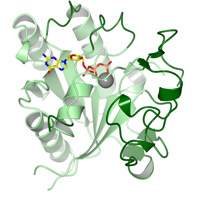
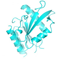
X-ray diffraction images
Data DOI: 10.15785/SBGRID/1147 | PDB ID 9MQ5: RCSB PDBe | Published: 1 Aug 2025
Boggon Laboratory, Yale University School of Medicine
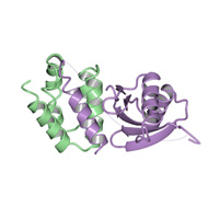
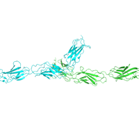
Native datasets
Data DOI: 10.15785/SBGRID/1107 | PDB ID 9BZ4: RCSB PDBe | Published: 4 Feb 2025
Boggon Laboratory, Yale University School of Medicine
4 sweeps
Data DOI: 10.15785/SBGRID/1010 | PDB ID 8GI4: RCSB PDBe | Published: 28 Nov 2023
Boggon Laboratory, Yale University School of Medicine
Four sweeps
Data DOI: 10.15785/SBGRID/879 | PDB ID 8DGQ: RCSB PDBe | Published: 28 Oct 2022
Boggon Laboratory, Yale University School of Medicine
Two sweeps, native
Data DOI: 10.15785/SBGRID/876 | PDB ID 7TPB: RCSB PDBe | Published: 28 Oct 2022
Boggon Laboratory, Yale University School of Medicine
Native data
Data DOI: 10.15785/SBGRID/854 | PDB ID 7S47: RCSB PDBe | Published: 18 Nov 2022
Boggon Laboratory, Yale University School of Medicine
Native data
Data DOI: 10.15785/SBGRID/853 | PDB ID 7S48: RCSB PDBe | Published: 18 Nov 2022
Boggon Laboratory, Yale University School of Medicine
Native datasets
Data DOI: 10.15785/SBGRID/852 | PDB ID 7S46: RCSB PDBe | Published: 18 Nov 2022
Boggon Laboratory, Yale University School of Medicine
Native dataset
Data DOI: 10.15785/SBGRID/782 | PDB ID 6WLY: RCSB PDBe | Published: 19 Jun 2020
Boggon Laboratory, Yale University School of Medicine
Native data
Data DOI: 10.15785/SBGRID/781 | PDB ID 6WLX: RCSB PDBe | Published: 19 Jun 2020
Boggon Laboratory, Yale University School of Medicine
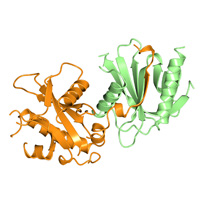
X-Ray Diffraction data from p120RasGAP C-terminal SH2 domain with p190RhoGAP pTyr-1087 peptide
Data DOI: 10.15785/SBGRID/775 | PDB ID 6WAY: RCSB PDBe | Published: 12 Jun 2020
Boggon Laboratory, Yale University School of Medicine
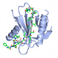
X-Ray Diffraction data from p120RasGAP C-terminal SH2 domain
Data DOI: 10.15785/SBGRID/774 | PDB ID 6WAX: RCSB PDBe | Published: 12 Jun 2020
Boggon Laboratory, Yale University School of Medicine
Native data. 3 passes.
Data DOI: 10.15785/SBGRID/699 | PDB ID 6PXC: RCSB PDBe | Published: 17 Dec 2019
Boggon Laboratory, Yale University School of Medicine
Native data. 2 passes.
Data DOI: 10.15785/SBGRID/698 | PDB ID 6PXB: RCSB PDBe | Published: 17 Dec 2019
Boggon Laboratory, Yale University School of Medicine
8 sweeps
Data DOI: 10.15785/SBGRID/575 | PDB ID 6D4G: RCSB PDBe | Published: 31 Aug 2018
Boggon Laboratory, Yale University School of Medicine
Native
Data DOI: 10.15785/SBGRID/563 | PDB ID 4Y5O: RCSB PDBe | Publication DOI: 10.1038/ncomms8937 | Published: 26 Jan 2018
Boggon Laboratory, Yale University School of Medicine
Native
Data DOI: 10.15785/SBGRID/562 | PDB ID 4WJ7: RCSB PDBe | Publication DOI: 10.1074/jbc.M114.616433 | Published: 26 Jan 2018
Boggon Laboratory, Yale University School of Medicine
Native
Data DOI: 10.15785/SBGRID/561 | PDB ID 4FQN: RCSB PDBe | Publication DOI: 10.1016/j.febslet.2012.12.011 | Published: 26 Jan 2018
Boggon Laboratory, Yale University School of Medicine
Native
Data DOI: 10.15785/SBGRID/558 | PDB ID 4TVQ: RCSB PDBe | Publication DOI: 10.1083/jcb.201407129 | Published: 26 Jan 2018
Boggon Laboratory, Yale University School of Medicine
Native
Data DOI: 10.15785/SBGRID/556 | PDB ID 4DXA: RCSB PDBe | Publication DOI: 10.1074/jbc.M112.361295 | Published: 26 Jan 2018
Boggon Laboratory, Yale University School of Medicine
Native
Data DOI: 10.15785/SBGRID/555 | PDB ID 3IXE: RCSB PDBe | Publication DOI: 10.1016/j.jsb.2009.12.002 | Published: 26 Jan 2018
Boggon Laboratory, Yale University School of Medicine
Iodide soak
Data DOI: 10.15785/SBGRID/554 | PDB ID 3F6Q: RCSB PDBe | Publication DOI: 10.1073/pnas.0811415106 | Published: 26 Jan 2018
Boggon Laboratory, Yale University School of Medicine
Native
Data DOI: 10.15785/SBGRID/553 | PDB ID 3ULR: RCSB PDBe | Publication DOI: 10.1107/S1744309111056132 | Published: 26 Jan 2018
Boggon Laboratory, Yale University School of Medicine
Native
Data DOI: 10.15785/SBGRID/552 | PDB ID 4EIH: RCSB PDBe | Publication DOI: 10.1074/jbc.M114.556480 | Published: 26 Jan 2018
Boggon Laboratory, Yale University School of Medicine
Se SAD
Data DOI: 10.15785/SBGRID/550 | PDB ID 4DX9: RCSB PDBe | Publication DOI: 10.1016/j.molcel.2012.12.005 | Published: 23 Jan 2018
Boggon Laboratory, Yale University School of Medicine
Native
Data DOI: 10.15785/SBGRID/549 | PDB ID 4DX8: RCSB PDBe | Publication DOI: 10.1016/j.molcel.2012.12.005 | Published: 23 Jan 2018
Boggon Laboratory, Yale University School of Medicine
Native
Data DOI: 10.15785/SBGRID/548 | PDB ID 4JIF: RCSB PDBe | Published: 23 Jan 2018
Boggon Laboratory, Yale University School of Medicine
Diffaction images
Data DOI: 10.15785/SBGRID/533 | PDB ID 5UPK: RCSB PDBe | Published: 22 Dec 2017
Boggon Laboratory, Yale University School of Medicine
Diffaction images
Data DOI: 10.15785/SBGRID/532 | PDB ID 5UPL: RCSB PDBe | Published: 22 Dec 2017
Boggon Laboratory, Yale University School of Medicine
360 images
Data DOI: 10.15785/SBGRID/514 | PDB ID 6BHC: RCSB PDBe | Published: 12 Dec 2017
Boggon Laboratory, Yale University School of Medicine
2 180 degree sweeps
Data DOI: 10.15785/SBGRID/494 | PDB ID 5VEF: RCSB PDBe | Published: 13 Oct 2017
Boggon Laboratory, Yale University School of Medicine
2 180 degree sweeps
Data DOI: 10.15785/SBGRID/493 | PDB ID 5VEE: RCSB PDBe | Published: 13 Oct 2017
Boggon Laboratory, Yale University School of Medicine
180 degrees
Data DOI: 10.15785/SBGRID/492 | PDB ID 5VED: RCSB PDBe | Published: 13 Oct 2017
Boggon Laboratory, Yale University School of Medicine
Native dataset
Data DOI: 10.15785/SBGRID/455 | PDB ID 5U4V: RCSB PDBe | Published: 12 Sep 2017
Boggon Laboratory, Yale University School of Medicine
Native datasets
Data DOI: 10.15785/SBGRID/454 | PDB ID 5U4U: RCSB PDBe | Published: 12 Sep 2017
Boggon Laboratory, Yale University School of Medicine
Selenomethionine labelled Tie2 FNIIIa–c collected at the remote wavelength
Data DOI: 10.15785/SBGRID/451 | PDB ID 5UTK: RCSB PDBe | Publication DOI: 10.1073/pnas.1617800114 | Published: 2 Jun 2017
Lemmon Laboratory, Yale University School of Medicine
X-ray diffraction
Data DOI: 10.15785/SBGRID/441 | PDB ID 4JDK: RCSB PDBe | Publication DOI: 10.1016/j.molcel.2013.11.013 | Published: 28 Mar 2017
Boggon Laboratory, Yale University School of Medicine
X-ray diffraction
Data DOI: 10.15785/SBGRID/440 | PDB ID 4JDJ: RCSB PDBe | Publication DOI: 10.1016/j.molcel.2013.11.013 | Published: 28 Mar 2017
Boggon Laboratory, Yale University School of Medicine
X-ray diffraction
Data DOI: 10.15785/SBGRID/439 | PDB ID 4JDH: RCSB PDBe | Publication DOI: 10.1016/j.molcel.2013.11.013 | Published: 28 Mar 2017
Boggon Laboratory, Yale University School of Medicine
X-ray diffraction
Data DOI: 10.15785/SBGRID/438 | PDB ID 4JDI: RCSB PDBe | Publication DOI: 10.1016/j.molcel.2013.11.013 | Published: 28 Mar 2017
Boggon Laboratory, Yale University School of Medicine
X-ray diffraction
Data DOI: 10.15785/SBGRID/437 | PDB ID 4FII: RCSB PDBe | Publication DOI: 10.1073/pnas.1214447109 | Published: 28 Mar 2017
Boggon Laboratory, Yale University School of Medicine
X-ray diffraction
Data DOI: 10.15785/SBGRID/436 | PDB ID 4FIH: RCSB PDBe | Publication DOI: 10.1073/pnas.1214447109 | Published: 28 Mar 2017
Boggon Laboratory, Yale University School of Medicine
X-ray diffraction
Data DOI: 10.15785/SBGRID/435 | PDB ID 4FIF: RCSB PDBe | Publication DOI: 10.1073/pnas.1214447109 | Published: 28 Mar 2017
Boggon Laboratory, Yale University School of Medicine
X-ray diffraction
Data DOI: 10.15785/SBGRID/434 | PDB ID 4FIG: RCSB PDBe | Publication DOI: 10.1073/pnas.1214447109 | Published: 28 Mar 2017
Boggon Laboratory, Yale University School of Medicine
X-ray diffraction
Data DOI: 10.15785/SBGRID/433 | PDB ID 4FIE: RCSB PDBe | Publication DOI: 10.1073/pnas.1214447109 | Published: 28 Mar 2017
Boggon Laboratory, Yale University School of Medicine
X-ray diffraction
Data DOI: 10.15785/SBGRID/432 | PDB ID 4FIJ: RCSB PDBe | Publication DOI: 10.1073/pnas.1214447109 | Published: 28 Mar 2017
Boggon Laboratory, Yale University School of Medicine
180°, 1° oscillations.
Data DOI: 10.15785/SBGRID/211 | PDB ID 5D68: RCSB PDBe | Publication DOI: 10.1016/j.jsb.2015.10.006 | Published: 5 Jan 2016
Boggon Laboratory, Yale University School of Medicine
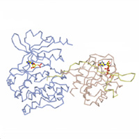
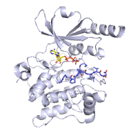
Crystal Structure of HIV-1 Reverse Transcriptase in Complex with 6-((4-((4-cyanophenyl)amino)-1,3,5-triazin-2-yl)amino)-5,7-dimethyl-2-naphthonitrile (JLJ639), a Non-nucleoside Inhibitor. 180 x 1 degree images collected for processing. Native data set collected on 24-ID-E at APS
Data DOI: 10.15785/SBGRID/168 | PDB ID 5C25: RCSB PDBe | Publication DOI: 10.1016/j.bmcl.2015.06.074 | Published: 8 Sep 2015
Anderson Laboratory, Yale University School of Medicine
native dataset
Data DOI: 10.15785/SBGRID/165 | PDB ID 4PGZ: RCSB PDBe | Publication DOI: 10.1016/j.molcel.2014.11.021 | Published: 6 Oct 2015
Schlessinger Laboratory, Yale University School of Medicine
native dataset
Data DOI: 10.15785/SBGRID/164 | PDB ID 4K94: RCSB PDBe | Publication DOI: 10.1073/pnas.1317118110 | Published: 6 Oct 2015
Schlessinger Laboratory, Yale University School of Medicine
Native data set
Data DOI: 10.15785/SBGRID/163 | PDB ID 4K9E: RCSB PDBe | Publication DOI: 10.1073/pnas.1317118110 | Published: 6 Oct 2015
Schlessinger Laboratory, Yale University School of Medicine
Crystal Structure of HIV-1 Reverse Transcriptase in Complex with (E)-3-(3-(4-chloro-2-(2-(2,4-dioxo-3,4-dihydropyrimidin-1(2H)-yl)ethoxy)phenoxy)phenyl)acrylonitrile (JLJ476), a non-nucleoside inhibitor. 180 x 1 degree images collected for processing. Native data set collected on X29A at NSLS-I.
Data DOI: 10.15785/SBGRID/19 | PDB ID 4LSL: RCSB PDBe | Publication DOI: 10.1111/cbdd.12266 | Published: 5 May 2015
Anderson Laboratory, Yale University School of Medicine
Native
Data DOI: 10.15785/SBGRID/5 | PDB ID 4TKN: RCSB PDBe | Publication DOI: 10.1074/jbc.M114.584011 | Published: 17 Apr 2015
Boggon Laboratory, Yale University School of Medicine