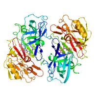
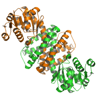
Engineered Rhizopuspepsin crystallized with a de novo designed modified peptide Ala-Cys-Val-Lys, with the Cys S and the Lys N covalently linked to a cyclohexal ring. Two molecules in the ASU.
Data DOI: 10.15785/SBGRID/989 | PDB ID 8FXQ: RCSB PDBe | Publication DOI: 10.1021/OL016090+ | Published: 3 Feb 2023
Rich Laboratory, University of Wisconsin-Madison
native data
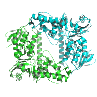
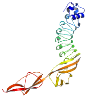
Data DOI: 10.15785/SBGRID/988 | PDB ID 6FRL: RCSB PDBe | Publication DOI: 10.1371/journal.pone.0196797 | Published: 3 Feb 2023
Niemann Laboratory, Bielefeld University
native data
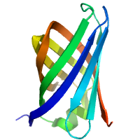
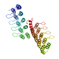
Data DOI: 10.15785/SBGRID/987 | PDB ID 7B1Z: RCSB PDBe | Publication DOI: 10.1107/S2053230X2100738X | Published: 7 Feb 2023
Niemann Laboratory, Bielefeld University
native data
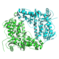
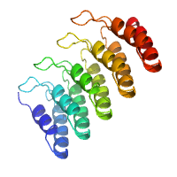
Data DOI: 10.15785/SBGRID/986 | PDB ID 6Y1W: RCSB PDBe | Publication DOI: 10.1107/S2059798320007731 | Published: 18 Jan 2023
Niemann Laboratory, Bielefeld University
native data
Data DOI: 10.15785/SBGRID/985 | PDB ID 6SMT: RCSB PDBe | Publication DOI: 10.3390/biom10081130 | Published: 17 Jan 2023
Niemann Laboratory, Bielefeld University
native data
Data DOI: 10.15785/SBGRID/984 | PDB ID 7NMS: RCSB PDBe | Publication DOI: 10.1107/S2059798322000432 | Published: 17 Jan 2023
Niemann Laboratory, Bielefeld University
native data
Data DOI: 10.15785/SBGRID/983 | PDB ID 7PV8: RCSB PDBe | Publication DOI: 10.1107/S2059798322000432 | Published: 17 Jan 2023
Niemann Laboratory, Bielefeld University
native data
Data DOI: 10.15785/SBGRID/982 | PDB ID 7PV9: RCSB PDBe | Publication DOI: 10.1107/S2059798322000432 | Published: 17 Jan 2023
Niemann Laboratory, Bielefeld University
native dataset
Data DOI: 10.15785/SBGRID/981 | PDB ID 8FRF: RCSB PDBe | Published: 9 Apr 2024
Garcia Laboratory, Stanford University
native dataset
Data DOI: 10.15785/SBGRID/980 | PDB ID 8FRE: RCSB PDBe | Published: 9 Apr 2024
Garcia Laboratory, Stanford University
(2S,3aS,8aS)-8a-hydroxy-3a-methyl-1,2,3,3a,8,8a-hexahydroindeno[2,1-b]pyrrole-2-carboxylic acid monohydrate
Data DOI: 10.15785/SBGRID/979 | Publication DOI: 10.1021/jacs.9b09864 | Published: 6 Jan 2023
Gonen Laboratory, University of California, Los Angeles
Ab initio structure of proteinase K from electron-counted MicroED data
Data DOI: 10.15785/SBGRID/978 | PDB ID 7SKX: RCSB PDBe | Publication DOI: 10.1038/s41592-022-01485-4 | Published: 3 Jan 2023
Gonen Laboratory, University of California, Los Angeles
MicroED structure of proteinase K from a platinum-coated, polished, single lamella at 1.79 Å resolution
Data DOI: 10.15785/SBGRID/977 | PDB ID 6PKR: RCSB PDBe | Publication DOI: 10.1016/j.str.2019.07.004 | Published: 3 Jan 2023
Gonen Laboratory, University of California, Los Angeles
High resolution dataset used in refinement
Data DOI: 10.15785/SBGRID/976 | PDB ID 8FFE: RCSB PDBe | Published: 3 Mar 2023
Garcia Laboratory, Stanford University
Native dataset collected from two crystals
Data DOI: 10.15785/SBGRID/975 | PDB ID 8FFE: RCSB PDBe | Published: 3 Mar 2023
Garcia Laboratory, Stanford University
native dataset
Data DOI: 10.15785/SBGRID/974 | PDB ID 8ENT: RCSB PDBe | Published: 23 Jun 2023
Garcia Laboratory, Stanford University
Native datasets
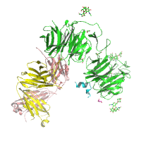
Data DOI: 10.15785/SBGRID/973 | PDB ID 8CUB: RCSB PDBe | Publication DOI: 10.1016/j.jmb.2022.167795 | Published: 13 Sep 2022
Lee Laboratory, University of Ottawa
Native datasets
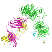
Data DOI: 10.15785/SBGRID/972 | PDB ID 8CUB: RCSB PDBe | Publication DOI: 10.1016/j.jmb.2022.167795 | Published: 13 Sep 2022
Lee Laboratory, University of Ottawa
Native datasets
Data DOI: 10.15785/SBGRID/971 | PDB ID 8CUB: RCSB PDBe | Publication DOI: 10.1016/j.jmb.2022.167795 | Published: 13 Sep 2022
Lee Laboratory, University of Ottawa
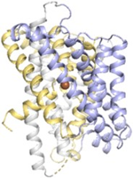
High resolution dataset collected from a single WT LCP crystal soaked in precipitant having cadmium.
Data DOI: 10.15785/SBGRID/969 | PDB ID 8E6M: RCSB PDBe | Published: 2 May 2023
Gaudet Laboratory, Harvard University