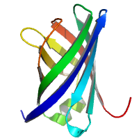
4 sweeps
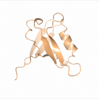
Data DOI: 10.15785/SBGRID/1010 | PDB ID 8GI4: RCSB PDBe | Published: 28 Nov 2023
Boggon Laboratory, Yale University School of Medicine
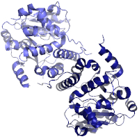
native data
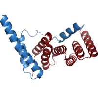
Data DOI: 10.15785/SBGRID/1009 | PDB ID 8ARB: RCSB PDBe | Published: 11 Apr 2023
Niemann Laboratory, Bielefeld University
Data with anomalous signal for Br (peak data set).
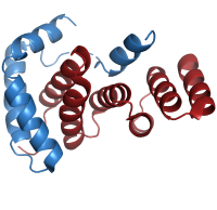
Data DOI: 10.15785/SBGRID/1008 | PDB ID 8ARC: RCSB PDBe | Published: 11 Apr 2023
Niemann Laboratory, Bielefeld University
native data
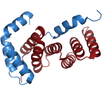
Data DOI: 10.15785/SBGRID/1007 | PDB ID 8ARA: RCSB PDBe | Published: 11 Apr 2023
Niemann Laboratory, Bielefeld University
data with anomalous signal for Br
Data DOI: 10.15785/SBGRID/1005 | PDB ID 8CJG: RCSB PDBe | Published: 16 Jun 2023
Niemann Laboratory, Bielefeld University
data with anomalous signal for Br
Data DOI: 10.15785/SBGRID/1004 | PDB ID 8CJF: RCSB PDBe | Published: 16 Jun 2023
Niemann Laboratory, Bielefeld University
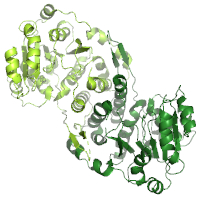
native data (almost perfectly non-merohedrally twinned)
Data DOI: 10.15785/SBGRID/1003 | PDB ID 8CJE: RCSB PDBe | Published: 16 Jun 2023
Niemann Laboratory, Bielefeld University
native data
Data DOI: 10.15785/SBGRID/1002 | PDB ID 8CJD: RCSB PDBe | Published: 16 Jun 2023
Niemann Laboratory, Bielefeld University
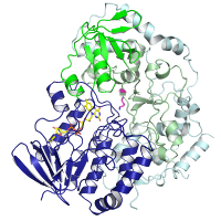
X-Ray Diffraction data from M. hassiacum GpgS, source of 7QOQ structure
Data DOI: 10.15785/SBGRID/1001 | PDB ID 7QOQ: RCSB PDBe | Published: 24 Feb 2023
Pereira Laboratory, IBMC/i3S, Universidade do Porto
X-Ray Diffraction data from M. hassiacum GpgS, source of 7PHO structure
Data DOI: 10.15785/SBGRID/1000 | PDB ID 7PHO: RCSB PDBe | Published: 24 Feb 2023
Pereira Laboratory, IBMC/i3S, Universidade do Porto
X-Ray Diffraction data from M. hassiacum GpgS, source of 7QIB structure
Data DOI: 10.15785/SBGRID/999 | PDB ID 7QIB: RCSB PDBe | Published: 24 Feb 2023
Pereira Laboratory, IBMC/i3S, Universidade do Porto
X-Ray Diffraction data from M. hassiacum GpgS, source of 7QI9 structure
Data DOI: 10.15785/SBGRID/998 | PDB ID 7QI9: RCSB PDBe | Published: 24 Feb 2023
Pereira Laboratory, IBMC/i3S, Universidade do Porto
X-Ray Diffraction data from M. hassiacum GpgS, source of 7PVL structure
Data DOI: 10.15785/SBGRID/997 | PDB ID 7PVL: RCSB PDBe | Published: 20 Mar 2026
Pereira Laboratory, IBMC/i3S, Universidade do Porto
X-Ray Diffraction data from M. hassiacum GpgS, source of 7QCP structure
Data DOI: 10.15785/SBGRID/996 | PDB ID 7QCP: RCSB PDBe | Published: 28 Feb 2023
Pereira Laboratory, IBMC/i3S, Universidade do Porto
X-Ray Diffraction data from M. hassiacum GpgS, source of 7P5L structure
Data DOI: 10.15785/SBGRID/995 | PDB ID 7P5L: RCSB PDBe | Published: 24 Feb 2023
Pereira Laboratory, IBMC/i3S, Universidade do Porto
X-Ray Diffraction data from M. hassiacum GpgS, source of 7PE4 structure
Data DOI: 10.15785/SBGRID/994 | PDB ID 7PE4: RCSB PDBe | Published: 28 Feb 2023
Pereira Laboratory, IBMC/i3S, Universidade do Porto
X-Ray Diffraction data from M. hassiacum GpgS, source of 7PD5 structure
Data DOI: 10.15785/SBGRID/993 | PDB ID 7PD5: RCSB PDBe | Published: 28 Feb 2023
Pereira Laboratory, IBMC/i3S, Universidade do Porto
X-Ray Diffraction data from M. hassiacum GpgS, source of 7PDO structure
Data DOI: 10.15785/SBGRID/992 | PDB ID 7PDO: RCSB PDBe | Published: 24 Feb 2023
Pereira Laboratory, IBMC/i3S, Universidade do Porto
X-Ray Diffraction data from M. hassiacum GpgS, source of 7P8G structure
Data DOI: 10.15785/SBGRID/991 | PDB ID 7P8G: RCSB PDBe | Published: 24 Feb 2023
Pereira Laboratory, IBMC/i3S, Universidade do Porto
native data
Data DOI: 10.15785/SBGRID/990 | PDB ID 4CV7: RCSB PDBe | Publication DOI: 10.1107/S2053230X14009911 | Published: 7 Feb 2023
Niemann Laboratory, Bielefeld University