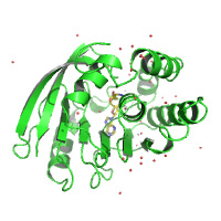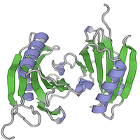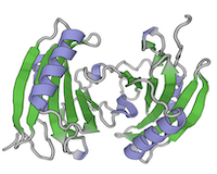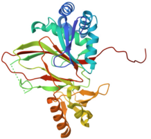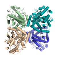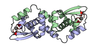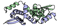
This contains datasets from two Mac1 crystals generated as part of Prism, Astera Institute’s effort to develop reproducible methods for generating high quality, diffuse scattering data. Each crystal has a corresponding background dataset in the subdirectory titled “background”. Similar crystals yielded the structure PDB 7KQO. Thus, no new PDB models were submitted. See README more details about the sample prep and beamtime.
Data DOI: 10.15785/SBGRID/1304 | Published: 16 Oct 2026
Prism, Astera Institute
This contains datasets from three Mac1 crystals generated as part of Prism, Astera Institute’s effort to develop reproducible methods for generating high quality, diffuse scattering data. Each crystal has a corresponding background dataset in the subdirectory titled “background”. Similar crystals yielded the structure PDB 7KQO. Thus, no new PDB models were submitted. See README more details about the sample prep and beamtime.
Data DOI: 10.15785/SBGRID/1303 | Published: 16 Oct 2026
Prism, Astera Institute
This contains datasets from three Mac1 crystals generated as part of Prism, Astera Institute’s effort to develop reproducible methods for generating high quality, diffuse scattering data. Each crystal has a corresponding background dataset in the subdirectory titled “background”. Similar crystals yielded the structure PDB 7KQO. Thus, no new PDB models were submitted. See README more details about the sample prep and beamtime.
Data DOI: 10.15785/SBGRID/1302 | Published: 13 Oct 2026
Ando Laboratory, Cornell University
This contains datasets from three Mac1 crystals generated as part of Prism, Astera Institute’s effort to develop reproducible methods for generating high quality, diffuse scattering data. Each crystal has a corresponding background dataset in the subdirectory titled “background”. Similar crystals yielded the structure PDB 7KQO. Thus, no new PDB models were submitted. See README more details about the sample prep and beamtime.
Data DOI: 10.15785/SBGRID/1301 | Published: 13 Oct 2026
Ando Laboratory, Cornell University
Native data of LpDot1 in complex with S-adenosyl homocysteine
Data DOI: 10.15785/SBGRID/1300 | Published: 12 Oct 2026
Buschiazzo Laboratory, Institut Pasteur de Montevideo
Room temperature Bragg and diffuse scattering data from Staphylococcal nuclease in complex with Ca2+ and thymidine-3'-5'-diphosphate (pdTp)
Data DOI: 10.15785/SBGRID/1295 | PDB ID 4WOR: RCSB PDBe | Publication DOI: 10.1073/pnas.94.12.6180 | Published: 25 Aug 2026
Prism, Astera Institute
X-ray diffraction data for crystals of DcrB-del37-GSSA-StrepII from Salmonella enterica at pH 4.0
Data DOI: 10.15785/SBGRID/1294 | PDB ID 13EE: RCSB PDBe | Published: 25 Aug 2026
May Laboratory, University of Wisconsin-La Crosse
X-ray diffraction data for crystals of DcrB-del37-GSSA-StrepII from Salmonella enterica at pH 5.0
Data DOI: 10.15785/SBGRID/1291 | PDB ID 13CZ: RCSB PDBe | Published: 14 Aug 2026
May Laboratory, University of Wisconsin-La Crosse
3 passes of 1440 raw diffraction images modeled by PDB ID 9B7C.
Data DOI: 10.15785/SBGRID/1276 | PDB ID 9B7C: RCSB PDBe | Publication DOI: 10.1126/sciadv.adj2921 | Published: 7 Jul 2026
Hekstra Laboratory, Harvard University
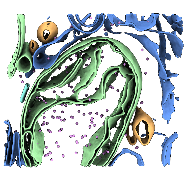
Two raw tilt-series (in MRC format) of FIB-milled G-TPP-treated HeLa cells that have been milled. The dataset contains mitochondria and cytoplasmic ribosomes as well as mHSP60-mHSP10 chaperonin "football" complexes. MRC files and MDOC files are provided. See the Processing Bundle below for all the scripts necessary to reconstruct the data to low-resolution following our tutorial. This tutorial involves template matching, subtomogram averaging, membrane segmentation, and particle place-back. See the processing instruction document for full details. Storage required: 4.4 G (dataset size) and 50G (storage for processing files when the tutorial has been completed).
Data DOI: 10.15785/SBGRID/1273 | Publication DOI: 10.1101/2025.10.06.680693 | Published: 10 Jul 2026
Ruben Fernandez-Busnadiego, University of Göttingen
X-ray diffraction images for Glutarate hydroxylase (GlaH) from Escherichia coli
Data DOI: 10.15785/SBGRID/1271 | PDB ID 8CDF: RCSB PDBe | Published: 7 Jul 2026
Schubert Laboratory, University of Pretoria
X-Ray Diffraction data from M. hassiacum GgH (Q434F mutant), source of 6G3N structure
Data DOI: 10.15785/SBGRID/1269 | PDB ID 6G3N: RCSB PDBe | Published: 8 May 2026
Pereira Laboratory, IBMC/i3S, Universidade do Porto
Structure of Human Spermidine Synthase in Complex with Putrescine
Data DOI: 10.15785/SBGRID/1268 | Published: 22 Sep 2026
Clinger Laboratory, Baylor University
293 K Structure of Human Spermidine Synthase
Data DOI: 10.15785/SBGRID/1266 | Published: 22 Sep 2026
Clinger Laboratory, Baylor University
273 K structure of Human Spermidine Synthase
Data DOI: 10.15785/SBGRID/1265 | Published: 22 Sep 2026
Clinger Laboratory, Baylor University
100 K structure of Human Spermidine synthase
Data DOI: 10.15785/SBGRID/1264 | Published: 22 Sep 2026
Clinger Laboratory, Baylor University
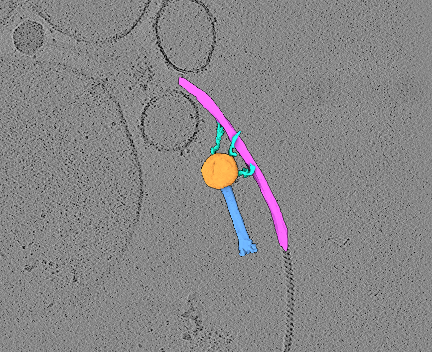
Two raw tilt-series (in MRC format) of highly-purified jumbo bacteriophage in association with bacterial flagella. MDOC files are also provided.
Data DOI: 10.15785/SBGRID/1263 | Publication DOI: doi.org/10.1038/s42003-025-09319-7 | Published: 21 Apr 2026
Briegel Laboratory, Institut Pasteur
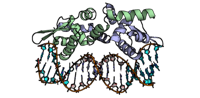
Crystal Structure of the Listeria monocytogenes CadC in complex with DNA
Data DOI: 10.15785/SBGRID/1261 | PDB ID 31NU: RCSB PDBe | Published: 28 Apr 2026
Cabanes Laboratory, i3S
Crystal Structure of the Listeria monocytogenes CadC with Cadmium
Data DOI: 10.15785/SBGRID/1260 | PDB ID 31RT: RCSB PDBe | Published: 28 Apr 2026
Cabanes Laboratory, i3S
Crystal Structure of the Listeria monocytogenes CadC
Data DOI: 10.15785/SBGRID/1259 | PDB ID 9T6R: RCSB PDBe | Published: 28 Apr 2026
Cabanes Laboratory, i3S