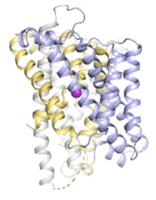
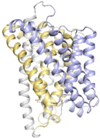
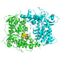
Datasets of Mn-bound D296A was collected from 3 LCP crystals. Image files that start C7run1_17 are from the first crystal, C7run2_19 are from the second crystal and C7run4_20 are from the third crystal.
Data DOI: 10.15785/SBGRID/968 | PDB ID 8E6L: RCSB PDBe | Published: 2 May 2023
Gaudet Laboratory, Harvard University
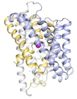
High resolution datasets of Mn-bound M230A mutant collected from 3 separate LCP crystals. Image files that start A131_run2 are from the first crystal, A132_run3 are from the second crystal and A133_run5 are from the third crystal.
Data DOI: 10.15785/SBGRID/967 | PDB ID 8E6I: RCSB PDBe | Published: 2 May 2023
Gaudet Laboratory, Harvard University
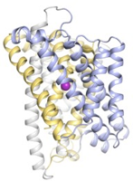
High resolution dataset of Mn-bound A47W mutant collected from a single LCP crystal
Data DOI: 10.15785/SBGRID/966 | PDB ID 8E6H: RCSB PDBe | Published: 2 May 2023
Gaudet Laboratory, Harvard University
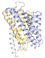
High resolution dataset of a single LCP crystal of WT soaked in precipitant containing manganese
Data DOI: 10.15785/SBGRID/964 | PDB ID 8E60: RCSB PDBe | Published: 2 May 2023
Gaudet Laboratory, Harvard University
High resolution dataset of a single LCP crystal of WT mock-soaked in precipitant
Data DOI: 10.15785/SBGRID/963 | PDB ID 8E5V: RCSB PDBe | Published: 2 May 2023
Gaudet Laboratory, Harvard University
High resolution dataset of WT protein obtained from single LCP crystal
Data DOI: 10.15785/SBGRID/962 | PDB ID 8E5S: RCSB PDBe | Published: 2 May 2023
Gaudet Laboratory, Harvard University
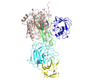
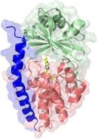
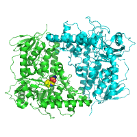
Fixed target serial crystallography data for PTP1B. A total of 6 individual chips (MiTeGen Sample supports) were collected. For each chip, a raster with an oscillation of 0.2 or 0.5 degree and 20um step. A total of 10 frames/chip with 1st 6 frames containing diffraction spots and the rest of the frames are blank. For more info on data collection and processing, please refer to this paper- https://doi.org/10.1101/2022.07.28.501725
Data DOI: 10.15785/SBGRID/961 | PDB ID 8DU7: RCSB PDBe | Publication DOI: 10.1101/2022.07.28.501725 | Published: 9 Sep 2022
Keedy Laboratory, Advanced Science Research Center, CUNY
Native dataset.
Data DOI: 10.15785/SBGRID/960 | PDB ID 7YZX: RCSB PDBe | Published: 9 Sep 2022
Cooney Laboratory, University of Limerick
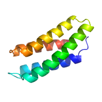
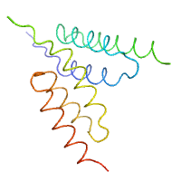
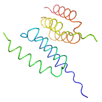
native dataset of neoleukin-4
Data DOI: 10.15785/SBGRID/959 | PDB ID 8DZ8: RCSB PDBe | Published: 24 Mar 2023
Garcia Laboratory, Stanford University
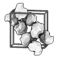
Hen lysozyme in orthorhombic space group at ambient temperature, diffuse scattering dataset
Data DOI: 10.15785/SBGRID/958 | PDB ID 8DZ7: RCSB PDBe | Published: 9 Sep 2022
Ando Laboratory, Cornell University
Hen lysozyme in tetragonal space group at ambient temperature, diffuse scattering dataset
Data DOI: 10.15785/SBGRID/957 | PDB ID 8DYZ: RCSB PDBe | Published: 9 Sep 2022
Ando Laboratory, Cornell University
3600 diffraction images
Data DOI: 10.15785/SBGRID/956 | PDB ID 8AQ8: RCSB PDBe | Published: 18 Jul 2023
Dohnalek Laboratory, Institute of Biotechnology of the Czech Academy of Sciences
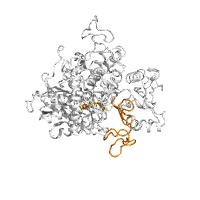
Crystal structure of human complex II assembly intermediate SDHA-SDHAF4
Data DOI: 10.15785/SBGRID/955 | PDB ID 8DYE: RCSB PDBe | Published: 20 Nov 2023
Iverson Laboratory, Vanderbilt University
Respiratory Complex II assembly intermediate SDHA-SDHAF2-SDHAF4
Data DOI: 10.15785/SBGRID/954 | PDB ID 8DYD: RCSB PDBe | Published: 21 Nov 2023
Iverson Laboratory, Vanderbilt University
native data
Data DOI: 10.15785/SBGRID/952 | PDB ID 8AD8: RCSB PDBe | Published: 21 Oct 2022
Niemann Laboratory, Bielefeld University
highly redundant data set
Data DOI: 10.15785/SBGRID/951 | PDB ID 8AD7: RCSB PDBe | Published: 21 Oct 2022
Niemann Laboratory, Bielefeld University
native dataset
Data DOI: 10.15785/SBGRID/929 | PDB ID 8DAC: RCSB PDBe | Published: 25 Jul 2023
Garcia Laboratory, Stanford University
native dataset
Data DOI: 10.15785/SBGRID/928 | PDB ID 8DAB: RCSB PDBe | Published: 25 Jul 2023
Garcia Laboratory, Stanford University
native dataset
Data DOI: 10.15785/SBGRID/927 | PDB ID 8DAA: RCSB PDBe | Published: 25 Jul 2023
Garcia Laboratory, Stanford University
native dataset
Data DOI: 10.15785/SBGRID/926 | PDB ID 8DA9: RCSB PDBe | Published: 25 Jul 2023
Garcia Laboratory, Stanford University