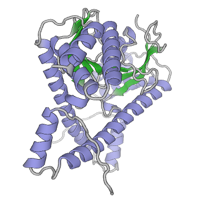
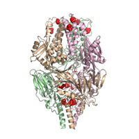
Iodine SAD dataset collected at 12670 electron volts. Crystal diffraction was measured at a temperature of 100 K using a 1 degree oscillation range.
Data DOI: 10.15785/SBGRID/198 | PDB ID 5CEG: RCSB PDBe | Publication DOI: 10.1016/j.cell.2015.09.055 | Published: 24 Nov 2015
Crosson Laboratory, University of Chicago
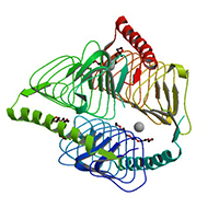
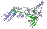
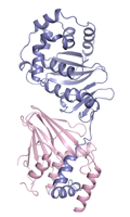
Diffraction data from this crystal of B. abortus RicA were collected at a temperature of 100 K using a 1° oscillation range.
Data DOI: 10.15785/SBGRID/197 | PDB ID 4N27: RCSB PDBe | Publication DOI: 10.1021/bi401373r | Published: 24 Nov 2015
Crosson Laboratory, University of Chicago
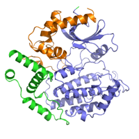
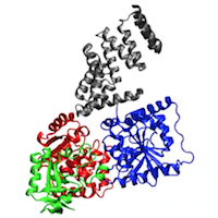
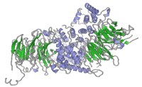
Native dataset for Hrr25:Mam1 complex, form 2
Data DOI: 10.15785/SBGRID/196 | PDB ID 5CZO: RCSB PDBe | Published: 9 Aug 2016
Corbett Laboratory, University of California, San Diego
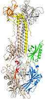
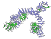
Native diffraction data for S. cerevisiae Hrr25:Mam1, form 1
Data DOI: 10.15785/SBGRID/195 | PDB ID 5CYZ: RCSB PDBe | Published: 9 Aug 2016
Corbett Laboratory, University of California, San Diego
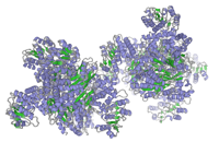
Native dataset
Data DOI: 10.15785/SBGRID/194 | PDB ID 5CXF: RCSB PDBe | Publication DOI: 10.1371/journal.ppat.1005227 | Published: 13 Nov 2015
Heldwein Laboratory, Tufts University School of Medicine
Native dataset
Data DOI: 10.15785/SBGRID/192 | PDB ID 5C6B: RCSB PDBe | Publication DOI: 10.1038/ncomms9143 | Published: 30 Oct 2015
McLellan Laboratory, University of Texas at Austin
Native dataset
Data DOI: 10.15785/SBGRID/191 | PDB ID 5C69: RCSB PDBe | Publication DOI: 10.1038/ncomms9143 | Published: 30 Oct 2015
McLellan Laboratory, University of Texas at Austin
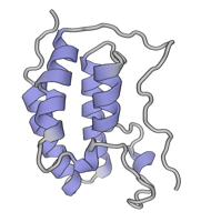
MD trajectory. The coordinates of the OGT–UDP–peptide complex (PDB 3PE4) were optimized in the Protein Preparation Wizard (Schrodinger 2009) where hydrogens were added; water molecules, UDP and peptide were stripped; and the structure was minimized using the OPLS2001 forcefield. The 1-μm simulation used the CHARM27 forcefield46, and the simple point charge model for water47. The CHARM27 forcefield was applied to the system using the VIPARR utility. The default Desmond relaxation was performed before simulation, and molecular dynamics were run at constant temperature (300 K) and pressure (1 bar). The simulation was performed by using the program Desmond, version 2.2.9.1.030 compiled by SBGrid on an optimized 64-node Linux-based InfiniBand cluster and took 75 days to complete. Molecular dynamics trajectories were processed and animated with VMD48.
Data DOI: 10.15785/SBGRID/190 | PDB ID 3PE4: RCSB PDBe | Publication DOI: 10.1038/nature09638 | Published: 3 Nov 2015
Sliz Laboratory, Harvard Medical School
Native dataset.
Data DOI: 10.15785/SBGRID/189 | PDB ID 4ZXS: RCSB PDBe | Publication DOI: 10.15252/embj.201592359 | Published: 30 Oct 2015
Heldwein Laboratory, Tufts University School of Medicine
Soluble portion of pseudorabies virus nuclear egress complex.
Data DOI: 10.15785/SBGRID/188 | PDB ID 4Z3U: RCSB PDBe | Publication DOI: 10.15252/embj.201592359 | Published: 30 Oct 2015
Heldwein Laboratory, Tufts University School of Medicine
mSlice Conditions: Waveform type : Linear X Galvo Offset, Interval (um), # of Pixels for Excitation (0) : 0 0.1 51 Z Galvo Offset, Interval (um), # of Pixels for Excitation (0) : -0.599257 0 251 Z PZT Offset, Interval (um), # of Pixels for Excitation (0) : 26 0 251 S PZT Offset, Interval (um), # of Pixels for Excitation (0) : 50 0.398406 251 # of stacks (0) : 200 Excitation Filter, Laser, Power (%), Exp(ms) (0) : N/A 488 20 40 Cycle lasers : per Z Z motion : Sample piezo
Data DOI: 10.15785/SBGRID/187 | Published: 16 Oct 2015
Kirchhausen Laboratory, Children's Hospital Boston
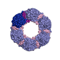
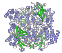
Bovine catalase microcrystals solved by MicroED
Data DOI: 10.15785/SBGRID/186 | PDB ID 3J7B: RCSB PDBe | Publication DOI: 10.7554/eLife.01345 | Published: 13 Oct 2015
Gonen Laboratory, University of California, Los Angeles
Native dataset
Data DOI: 10.15785/SBGRID/184 | Publication DOI: 10.1021/jm501120z | Published: 23 Oct 2015
Blacklow Laboratory, Harvard Medical School
SeMet data, collected on two separate spots on a single crystal
Data DOI: 10.15785/SBGRID/182 | PDB ID 4FHM: RCSB PDBe | Publication DOI: 10.1073/pnas.1205151109 | Published: 6 Oct 2015
Schwartz Laboratory, Massachusetts Institute of Technology
native data, collected on four spots on the same crystal
Data DOI: 10.15785/SBGRID/181 | PDB ID 4FHM: RCSB PDBe | Publication DOI: 10.1073/pnas.1205151109 | Published: 6 Oct 2015
Schwartz Laboratory, Massachusetts Institute of Technology
Ta6Br12 derivative data
Data DOI: 10.15785/SBGRID/180 | PDB ID 4FHN: RCSB PDBe | Publication DOI: 10.1073/pnas.1205151109 | Published: 6 Oct 2015
Schwartz Laboratory, Massachusetts Institute of Technology
native data, collected in two separate spots
Data DOI: 10.15785/SBGRID/179 | PDB ID 4FHN: RCSB PDBe | Publication DOI: 10.1073/pnas.1205151109 | Published: 6 Oct 2015
Schwartz Laboratory, Massachusetts Institute of Technology
native data
Data DOI: 10.15785/SBGRID/178 | PDB ID 4FCC: RCSB PDBe | Publication DOI: 10.1073/pnas.1205151109 | Published: 6 Oct 2015
Schwartz Laboratory, Massachusetts Institute of Technology