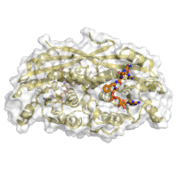
native dataset
Data DOI: 10.15785/SBGRID/881 | PDB ID 7RAA: RCSB PDBe | Published: 15 Mar 2022
Garcia Laboratory, Stanford University
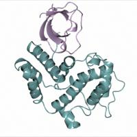
Native dataset. Phased by molecular replacement with PDB 6QAJ.
Data DOI: 10.15785/SBGRID/880 | PDB ID 7Z36: RCSB PDBe | Published: 21 Oct 2022
Modis Laboratory, University of Cambridge
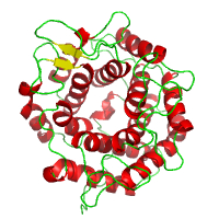
Four sweeps
Data DOI: 10.15785/SBGRID/879 | PDB ID 8DGQ: RCSB PDBe | Published: 28 Oct 2022
Boggon Laboratory, Yale University School of Medicine
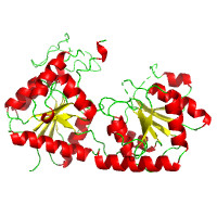
Native dataset to 1.77A
Data DOI: 10.15785/SBGRID/878 | PDB ID 7TSX: RCSB PDBe | Published: 6 Jan 2023
Corbett Laboratory, University of California, San Diego
Native dataset to 2.11 A resolution
Data DOI: 10.15785/SBGRID/877 | PDB ID 7TSQ: RCSB PDBe | Published: 6 Jan 2023
Corbett Laboratory, University of California, San Diego
Two sweeps, native
Data DOI: 10.15785/SBGRID/876 | PDB ID 7TPB: RCSB PDBe | Published: 28 Oct 2022
Boggon Laboratory, Yale University School of Medicine
X-Ray Diffraction data from M. hassiacum MmpH (Se-Met data set)
Data DOI: 10.15785/SBGRID/875 | Published: 24 Jan 2023
Pereira Laboratory, IBMC/i3S, Universidade do Porto
X-Ray Diffraction data from M. hassiacum MmpH, source of 7QSJ structure (native data set)
Data DOI: 10.15785/SBGRID/874 | PDB ID 7QSJ: RCSB PDBe | Published: 24 Jan 2023
Pereira Laboratory, IBMC/i3S, Universidade do Porto
X-Ray Diffraction data from M. hassiacum ManT, source of 7QSG structure
Data DOI: 10.15785/SBGRID/873 | PDB ID 7QSG: RCSB PDBe | Published: 24 Jan 2023
Pereira Laboratory, IBMC/i3S, Universidade do Porto
Native data
Data DOI: 10.15785/SBGRID/868 | PDB ID 7T5V: RCSB PDBe | Published: 9 Sep 2022
Corbett Laboratory, University of California, San Diego
Native data
Data DOI: 10.15785/SBGRID/867 | PDB ID 7T5W: RCSB PDBe | Published: 9 Sep 2022
Corbett Laboratory, University of California, San Diego
Native data
Data DOI: 10.15785/SBGRID/866 | PDB ID 7T5U: RCSB PDBe | Published: 9 Sep 2022
Corbett Laboratory, University of California, San Diego
Selenomethione SAD (peak) data
Data DOI: 10.15785/SBGRID/865 | PDB ID 7T5T: RCSB PDBe | Published: 9 Sep 2022
Corbett Laboratory, University of California, San Diego
Native data
Data DOI: 10.15785/SBGRID/864 | PDB ID 7T5T: RCSB PDBe | Published: 9 Sep 2022
Corbett Laboratory, University of California, San Diego
X-Ray Diffraction data from Candida albicans Ras1 guanine-nucleotide exchange factor, low resolution dataset, crystal 2
Data DOI: 10.15785/SBGRID/861 | Published: 25 Jul 2023
Pereira Laboratory, IBMC/i3S, Universidade do Porto
X-Ray Diffraction data from Candida albicans Ras1 guanine-nucleotide exchange factor, low resolution dataset, crystal 1
Data DOI: 10.15785/SBGRID/860 | Published: 25 Jul 2023
Pereira Laboratory, IBMC/i3S, Universidade do Porto
X-Ray Diffraction data from Candida albicans Ras1 guanine-nucleotide exchange factor, source of 7NZZ structure
Data DOI: 10.15785/SBGRID/859 | PDB ID 7NZZ: RCSB PDBe | Published: 25 Jul 2023
Pereira Laboratory, IBMC/i3S, Universidade do Porto
native dataset, P4222 crystal form
Data DOI: 10.15785/SBGRID/858 | PDB ID 7R85: RCSB PDBe | Published: 9 Nov 2021
Garcia Laboratory, Stanford University
native dataset, P21 crystal form
Data DOI: 10.15785/SBGRID/857 | PDB ID 7R84: RCSB PDBe | Published: 9 Nov 2021
Garcia Laboratory, Stanford University
native dataset
Data DOI: 10.15785/SBGRID/856 | PDB ID 7R86: RCSB PDBe | Published: 9 Nov 2021
Garcia Laboratory, Stanford University