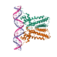
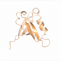
Native dataset
Data DOI: 10.15785/SBGRID/1075 | PDB ID 6NSM: RCSB PDBe | Publication DOI: https://doi.org/10.1101/2024.01.16.572601 | Published: 26 Jan 2024
Madden Laboratory, Dartmouth Geisel School of Medicine
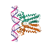
Native dataset
Data DOI: 10.15785/SBGRID/1074 | PDB ID 6NSN: RCSB PDBe | Published: 26 Jan 2024
Madden Laboratory, Dartmouth Geisel School of Medicine
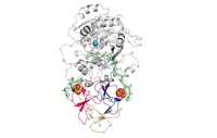
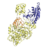
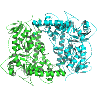
Native data set to 2.3 angstrom resolution.
Data DOI: 10.15785/SBGRID/1071 | PDB ID 8V9Q: RCSB PDBe | Published: 19 Feb 2024
Samara Laboratory, NIH
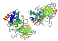
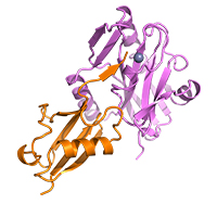
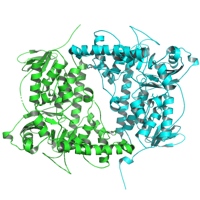
Apo WT Protein tyrosine phosphatase 1B crystal shot at cryogenic temp at the NSLS-II NYX beamline
Data DOI: 10.15785/SBGRID/1060 | PDB ID 8U1E: RCSB PDBe | Publication DOI: https://doi.org/10.1101/2023.09.12.557361 | Published: 21 Nov 2023
Keedy Laboratory, Advanced Science Research Center, CUNY
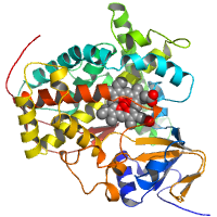
Native dataset, Cu-bound protein
Data DOI: 10.15785/SBGRID/1054 | PDB ID 8UM6: RCSB PDBe | Published: 7 Feb 2024
Fisher Laboratory, Lehigh University
Single-wavelength dataset
Data DOI: 10.15785/SBGRID/1042 | PDB ID 8TYZ: RCSB PDBe | Published: 9 Jul 2024
Corbett Laboratory, University of California, San Diego
Single-wavelength dataset
Data DOI: 10.15785/SBGRID/1041 | PDB ID 8TZ0: RCSB PDBe | Published: 9 Jul 2024
Corbett Laboratory, University of California, San Diego
Single-wavelength dataset
Data DOI: 10.15785/SBGRID/1040 | PDB ID 8TYY: RCSB PDBe | Published: 9 Jul 2024
Corbett Laboratory, University of California, San Diego
Single-wavelength dataset
Data DOI: 10.15785/SBGRID/1039 | PDB ID 8TYX: RCSB PDBe | Published: 9 Jul 2024
Corbett Laboratory, University of California, San Diego
native data
Data DOI: 10.15785/SBGRID/1037 | PDB ID 8Q2F: RCSB PDBe | Published: 4 Feb 2025
Niemann Laboratory, Bielefeld University
Two datasets collected on the same crystal.
Data DOI: 10.15785/SBGRID/1035 | PDB ID 8BFY: RCSB PDBe | Published: 18 Jul 2023
Kerff Laboratory, University of Liège
SeMet dataset
Data DOI: 10.15785/SBGRID/1034 | PDB ID 8TCH: RCSB PDBe | Published: 20 Mar 2026
Rice Laboratory, University of Chicago
Native dataset
Data DOI: 10.15785/SBGRID/1033 | PDB ID 8TCH: RCSB PDBe | Publication DOI: NA | Published: 20 Mar 2026
Rice Laboratory, University of Chicago
native data set
Data DOI: 10.15785/SBGRID/1032 | PDB ID 8T85: RCSB PDBe | Published: 2 Feb 2024
Deaconescu Laboratory, Brown University
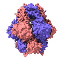
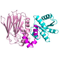
homo dimer with peptide in between
Data DOI: 10.15785/SBGRID/1026 | PDB ID 7QSA: RCSB PDBe | Publication DOI: 10.1042/BCJ20220037 | Published: 16 May 2023
Kvansakul Laboratory, La Trobe University
native data
Data DOI: 10.15785/SBGRID/1023 | PDB ID 7AQV: RCSB PDBe | Publication DOI: 10.3390/cryst10121135 | Published: 25 Apr 2023
Niemann Laboratory, Bielefeld University
native data
Data DOI: 10.15785/SBGRID/1022 | PDB ID 7AQU: RCSB PDBe | Publication DOI: 10.3390/cryst10121135 | Published: 25 Apr 2023
Niemann Laboratory, Bielefeld University
4 sweeps
Data DOI: 10.15785/SBGRID/1010 | PDB ID 8GI4: RCSB PDBe | Published: 28 Nov 2023
Boggon Laboratory, Yale University School of Medicine
native data
Data DOI: 10.15785/SBGRID/1009 | PDB ID 8ARB: RCSB PDBe | Published: 11 Apr 2023
Niemann Laboratory, Bielefeld University
Data with anomalous signal for Br (peak data set).
Data DOI: 10.15785/SBGRID/1008 | PDB ID 8ARC: RCSB PDBe | Published: 11 Apr 2023
Niemann Laboratory, Bielefeld University