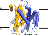
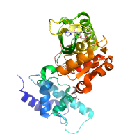
Native dataset.
Data DOI: 10.15785/SBGRID/335 | Publication DOI: 10.1016/j.str.2016.09.017 | Published: 15 Nov 2016
Gaudet Laboratory, Harvard University
The second of two osmium containing data sets.
Data DOI: 10.15785/SBGRID/334 | PDB ID 5KTE: RCSB PDBe | Publication DOI: 10.1016/j.str.2016.09.017 | Published: 15 Nov 2016
Gaudet Laboratory, Harvard University
One of two Osmium containing data sets.
Data DOI: 10.15785/SBGRID/333 | PDB ID 5KTE: RCSB PDBe | Publication DOI: 10.1016/j.str.2016.09.017 | Published: 15 Nov 2016
Gaudet Laboratory, Harvard University
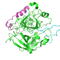
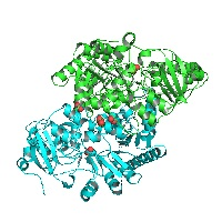
Native dataset.
Data DOI: 10.15785/SBGRID/332 | PDB ID 5KTE: RCSB PDBe | Publication DOI: 10.1016/j.str.2016.09.017 | Published: 15 Nov 2016
Gaudet Laboratory, Harvard University
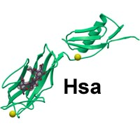
Hsa bound to the sialyl-T antigen trisaccharide
Data DOI: 10.15785/SBGRID/329 | Published: 4 Jan 2022
Iverson Laboratory, Vanderbilt University
Crystals of the Hsa binding region
Data DOI: 10.15785/SBGRID/328 | Published: 4 Jan 2022
Iverson Laboratory, Vanderbilt University
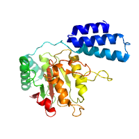
Native dataset for S. cerevisiae Csm1:Ulp2 complex
Data DOI: 10.15785/SBGRID/327 | PDB ID 5V1A: RCSB PDBe | Published: 9 May 2017
Corbett Laboratory, University of California, San Diego
Native data
Data DOI: 10.15785/SBGRID/325 | PDB ID 5K8R: RCSB PDBe | Published: 29 Jul 2016
Gaudet Laboratory, Harvard University
X-ray Diffraction data from Human thrombin in complex with a disulfated madanin-1 fragment, source of 5L6N structure
Data DOI: 10.15785/SBGRID/324 | PDB ID 5L6N: RCSB PDBe | Published: 24 Mar 2017
Pereira Laboratory, IBMC/i3S, Universidade do Porto
Native dataset
Data DOI: 10.15785/SBGRID/320 | PDB ID 4YPN: RCSB PDBe | Publication DOI: 10.1016/j.str.2016.03.001 | Published: 31 May 2016
Chang Laboratory, Academia Sinica
Native dataset
Data DOI: 10.15785/SBGRID/319 | PDB ID 4YPL: RCSB PDBe | Publication DOI: 10.1016/j.str.2016.03.001 | Published: 31 May 2016
Chang Laboratory, Academia Sinica
Native dataset
Data DOI: 10.15785/SBGRID/316 | PDB ID 5E7S: RCSB PDBe | Publication DOI: 10.1016/j.str.2016.03.003 | Published: 31 May 2016
Chang Laboratory, Academia Sinica
Native dataset
Data DOI: 10.15785/SBGRID/314 | PDB ID 4YPM: RCSB PDBe | Publication DOI: 10.1016/j.str.2016.03.003 | Published: 31 May 2016
Chang Laboratory, Academia Sinica
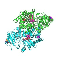
Succinyl-CoA:acetate CoA-transferase (AarCH6) bound to acetate and degradation products from the acetyl-CoA analogue dethiaacetyl-CoA (AcMX)
Data DOI: 10.15785/SBGRID/311 | PDB ID 5E5H: RCSB PDBe | Publication DOI: 10.3389/fchem.2016.00023 | Published: 31 May 2016
Kappock Laboratory, Purdue University
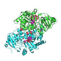
Succinyl-CoA:acetate CoA-transferase (AarCH6) bound to acetate and the CoA analogue 3'-phosphoadenosine 5'-(O-(N-propyl-R-pantothenamide))pyrophosphate (MX)
Data DOI: 10.15785/SBGRID/310 | PDB ID 5DW6: RCSB PDBe | Publication DOI: 10.3389/fchem.2016.00023 | Published: 31 May 2016
Kappock Laboratory, Purdue University
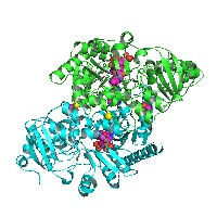
Succinyl-CoA:acetate CoA-transferase (AarCH6) bound to the CoA analogue 3'-phosphoadenosine 5'-(O-(N-propylpantothenamide))pyrophosphate (MX)
Data DOI: 10.15785/SBGRID/309 | PDB ID 5DW5: RCSB PDBe | Publication DOI: 10.3389/fchem.2016.00023 | Published: 31 May 2016
Kappock Laboratory, Purdue University
Succinyl-CoA:acetate CoA-transferase (AarCH6) bound to acetate
Data DOI: 10.15785/SBGRID/308 | PDB ID 5DW4: RCSB PDBe | Publication DOI: 10.3389/fchem.2016.00023 | Published: 31 May 2016
Kappock Laboratory, Purdue University
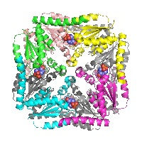
Crystal structure of Treponema denticola PurE, a 5-aminoimidazole ribonucleotide (AIR) carboxylase, bound to AIR
Data DOI: 10.15785/SBGRID/302 | PDB ID 3RGG: RCSB PDBe | Publication DOI: 10.1021/bi102033a | Published: 31 May 2016
Kappock Laboratory, Purdue University
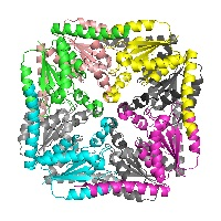
Crystal structure of Treponema denticola PurE, a 5-aminoimidazole ribonucleotide (AIR) carboxylase
Data DOI: 10.15785/SBGRID/301 | PDB ID 3RG8: RCSB PDBe | Publication DOI: 10.1021/bi102033a | Published: 8 Jul 2016
Kappock Laboratory, Purdue University
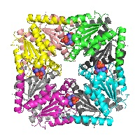
Structure of PurE (N5-carboxyaminoimidazole ribonucleotide mutase) H59N from the acidophilic bacterium Acetobacter aceti, bound to isocair
Data DOI: 10.15785/SBGRID/300 | PDB ID 2FWP: RCSB PDBe | Publication DOI: 10.1021/bi060465n | Published: 8 Jul 2016
Kappock Laboratory, Purdue University