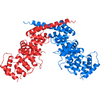
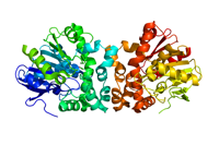
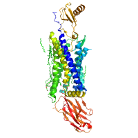
Native dataset to 2.6 A resolution of Human Annexin A13a
Data DOI: 10.15785/SBGRID/353 | PDB ID 6B3I: RCSB PDBe | Published: 21 Dec 2018
Iverson Laboratory, Vanderbilt University
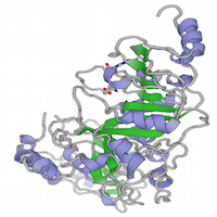
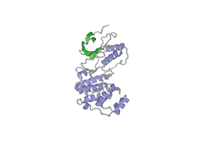
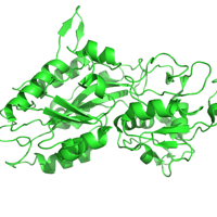
Fluoroacetate dehalogenase Apo with no Halides
Data DOI: 10.15785/SBGRID/352 | Published: 16 Aug 2016
Pai Laboratory, University of Toronto
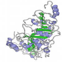
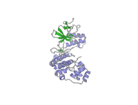
Fluoroacetate dehalogenase H280N with covalent intermediate in both subunits
Data DOI: 10.15785/SBGRID/351 | Published: 16 Aug 2016
Pai Laboratory, University of Toronto
Fluoroacetate dehalogenase H280N with covalent intermediate in 1 subunit
Data DOI: 10.15785/SBGRID/350 | Published: 16 Aug 2016
Pai Laboratory, University of Toronto
Native dataset
Data DOI: 10.15785/SBGRID/349 | PDB ID 5K7W: RCSB PDBe | Publication DOI: 10.1016/j.molcel.2016.05.041 | Published: 5 Aug 2016
Nam Laboratory, UT Southwestern Medical Center
Native dataset
Data DOI: 10.15785/SBGRID/348 | PDB ID 5K7U: RCSB PDBe | Publication DOI: 10.1016/j.molcel.2016.05.041 | Published: 5 Aug 2016
Nam Laboratory, UT Southwestern Medical Center
Native dataset
Data DOI: 10.15785/SBGRID/346 | PDB ID 5K7M: RCSB PDBe | Publication DOI: 10.1016/j.molcel.2016.05.041 | Published: 5 Aug 2016
Nam Laboratory, UT Southwestern Medical Center
Fluoroacetate dehalogenase cocrystallized with glycolate
Data DOI: 10.15785/SBGRID/345 | Published: 16 Aug 2016
Pai Laboratory, University of Toronto
Fluoroacetate dehalogenase cocrystallized with chloroacetate
Data DOI: 10.15785/SBGRID/344 | Published: 16 Aug 2016
Pai Laboratory, University of Toronto
Fluoroacetate dehalogenase with 5'-F-tryptophans
Data DOI: 10.15785/SBGRID/343 | Published: 16 Aug 2016
Pai Laboratory, University of Toronto
Native data set
Data DOI: 10.15785/SBGRID/342 | PDB ID 5EKO: RCSB PDBe | Publication DOI: 10.1016/j.bbagen.2016.06.023 | Published: 5 Aug 2016
Brett Laboratory, Washington U. School of Medicine
Native data set
Data DOI: 10.15785/SBGRID/341 | PDB ID 5EKN: RCSB PDBe | Publication DOI: 10.1016/j.bbagen.2016.06.023 | Published: 5 Aug 2016
Brett Laboratory, Washington U. School of Medicine
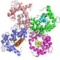
native dataset
Data DOI: 10.15785/SBGRID/340 | PDB ID 5IWL: RCSB PDBe | Publication DOI: 10.1172/JCI81603 | Published: 2 Aug 2016
Garcia Laboratory, Stanford University
native datasets
Data DOI: 10.15785/SBGRID/339 | PDB ID 4XT1: RCSB PDBe | Publication DOI: 10.1126/science.aaa5026 | Published: 2 Aug 2016
Garcia Laboratory, Stanford University
These are twinned datasets which were successfully merged together and solved with MR-SAD.
Data DOI: 10.15785/SBGRID/338 | PDB ID 5DOW: RCSB PDBe | Published: 29 Jul 2016
Looger Laboratory, HHMI/Janelia Research Campus
Native dataset
Data DOI: 10.15785/SBGRID/337 | PDB ID 5J1D: RCSB PDBe | Published: 29 Jul 2016
Murthy Laboratory, Indian Institute of Science
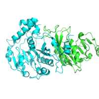
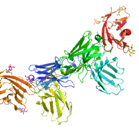
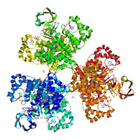
Acetobacter aceti citrate synthase (AarA), a type II citrate si-synthase (EC 2.3.3.1) insensitive to NADH, complexed with oxaloacetate and carboxymethyldethia coenzyme A (CMX)
Data DOI: 10.15785/SBGRID/336 | PDB ID 2H12: RCSB PDBe | Publication DOI: 10.1021/bi061083k | Published: 12 Jul 2016
Kappock Laboratory, Purdue University
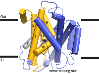
Native dataset.
Data DOI: 10.15785/SBGRID/335 | Publication DOI: 10.1016/j.str.2016.09.017 | Published: 15 Nov 2016
Gaudet Laboratory, Harvard University
The second of two osmium containing data sets.
Data DOI: 10.15785/SBGRID/334 | PDB ID 5KTE: RCSB PDBe | Publication DOI: 10.1016/j.str.2016.09.017 | Published: 15 Nov 2016
Gaudet Laboratory, Harvard University
One of two Osmium containing data sets.
Data DOI: 10.15785/SBGRID/333 | PDB ID 5KTE: RCSB PDBe | Publication DOI: 10.1016/j.str.2016.09.017 | Published: 15 Nov 2016
Gaudet Laboratory, Harvard University