Native dataset
Data DOI: 10.15785/SBGRID/376 | PDB ID 5SZN: RCSB PDBe | Publication DOI: 10.7554/eLife.20930 | Published: 1 Nov 2016
Shapiro Laboratory, Columbia University
Native dataset
Data DOI: 10.15785/SBGRID/375 | PDB ID 5SZM: RCSB PDBe | Publication DOI: 10.7554/eLife.20930 | Published: 1 Nov 2016
Shapiro Laboratory, Columbia University
Native dataset
Data DOI: 10.15785/SBGRID/374 | PDB ID 5SZL: RCSB PDBe | Publication DOI: 10.7554/eLife.20930 | Published: 1 Nov 2016
Shapiro Laboratory, Columbia University
Native dataset
Data DOI: 10.15785/SBGRID/373 | PDB ID 5K70: RCSB PDBe | Published: 28 Oct 2016
Shapiro Laboratory, Columbia University
Native dataset
Data DOI: 10.15785/SBGRID/372 | PDB ID 5K6Z: RCSB PDBe | Published: 28 Oct 2016
Shapiro Laboratory, Columbia University
Native dataset
Data DOI: 10.15785/SBGRID/371 | PDB ID 5K6Y: RCSB PDBe | Publication DOI: 10.7554/eLife.19058 | Published: 28 Oct 2016
Shapiro Laboratory, Columbia University
Native dataset
Data DOI: 10.15785/SBGRID/370 | PDB ID 5K6X: RCSB PDBe | Publication DOI: 10.7554/eLife.19058 | Published: 28 Oct 2016
Shapiro Laboratory, Columbia University
Native dataset
Data DOI: 10.15785/SBGRID/369 | PDB ID 5K6W: RCSB PDBe | Publication DOI: 10.7554/eLife.19058 | Published: 28 Oct 2016
Shapiro Laboratory, Columbia University
The XFEL data were collected at the X-ray Pump Probe (XPP) endstation of the Linac Coherent Light Source (LCLS) at the SLAC National Accelerator Laboratory, using a goniometer-based fixed target sample delivery station and an automatic sample loading system. We used a 30-micron XFEL beam with a pulse duration of 40 fs in SASE mode. A total of 148 crystals were screened, yielding 789 images with usable diffraction.
Data DOI: 10.15785/SBGRID/365 | PDB ID 5KJ7: RCSB PDBe | Published: 28 Oct 2016
Brunger Laboratory, Stanford University
native x-ray diffraction dataset
Data DOI: 10.15785/SBGRID/364 | PDB ID 5TEU: RCSB PDBe | Published: 23 Sep 2016
Crosson Laboratory, University of Chicago
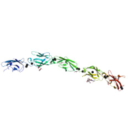
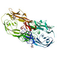
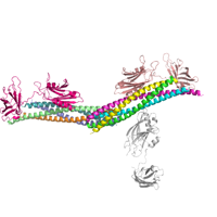
Native dataset used to determine the structure of Human Protocadherin-15 EC3-5 D414A Variant using molecular replacement.
Data DOI: 10.15785/SBGRID/363 | PDB ID 5T4N: RCSB PDBe | Published: 14 Feb 2017
Gaudet Laboratory, Harvard University
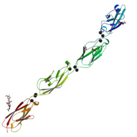
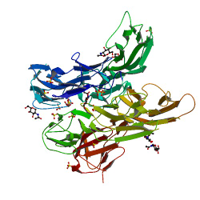
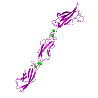
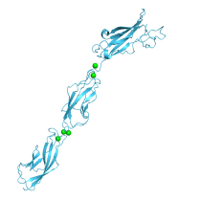
Native dataset used to determine low resolution structure of Mouse Protocadherin-15 EC4-5. The EC4-5 structure was then used as a molecular replacement search model to determine the structure of Human Protocadherin-15 EC3-5 (PDB Code: 5T4M)
Data DOI: 10.15785/SBGRID/362 | PDB ID 5T4M: RCSB PDBe | Published: 14 Feb 2017
Gaudet Laboratory, Harvard University
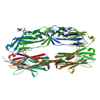
Native dataset of Human Protocadherin-15 EC3-5 used to determine structure using molecular replacement.
Data DOI: 10.15785/SBGRID/361 | PDB ID 5T4M: RCSB PDBe | Published: 14 Feb 2017
Gaudet Laboratory, Harvard University
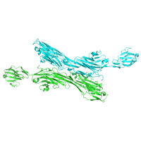
Native dataset
Data DOI: 10.15785/SBGRID/360 | PDB ID 5L1X: RCSB PDBe | Published: 30 Aug 2016
McLellan Laboratory, University of Texas at Austin
Native data for PDB entry 5SZC
Data DOI: 10.15785/SBGRID/359 | PDB ID 5SZC: RCSB PDBe | Published: 5 Sep 2017
Corbett Laboratory, University of California, San Diego
Native data for PDB 5SZB
Data DOI: 10.15785/SBGRID/358 | PDB ID 5SZB: RCSB PDBe | Published: 5 Sep 2017
Corbett Laboratory, University of California, San Diego
Zinc SAD data for PDB entry 5SZB
Data DOI: 10.15785/SBGRID/357 | PDB ID 5SZB: RCSB PDBe | Published: 5 Sep 2017
Corbett Laboratory, University of California, San Diego
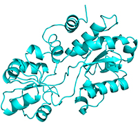
X-ray diffraction data for the methyltransferase domain of EvdMO1 (EvdMdO1) to 1.10 A resolution. Data were collected from a sample co-crystallized in the presence of 2 mM SAH and 10 mM fucose. No density for the fucose is observed, but an active site loop becomes disordered in the presence of fucose.
Data DOI: 10.15785/SBGRID/356 | Published: 20 Nov 2018
Iverson Laboratory, Vanderbilt University
X-ray diffraction data for the methyltransferase domain of EvdMO1 (EvdMdO1) to 1.15 A resolution.
Data DOI: 10.15785/SBGRID/355 | Published: 19 Nov 2018
Iverson Laboratory, Vanderbilt University
X-ray diffraction data to 3.35 A resolution for the full-length EvdMO1 enzyme
Data DOI: 10.15785/SBGRID/354 | Published: 20 Nov 2018
Iverson Laboratory, Vanderbilt University