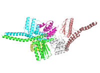
Native dataset
Data DOI: 10.15785/SBGRID/404 | PDB ID 5EHS: RCSB PDBe | Publication DOI: 10.1016/j.neuron.2016.10.058 | Published: 13 Dec 2016
Mayer Laboratory, National Institutes of Health
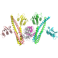
Native dataset
Data DOI: 10.15785/SBGRID/403 | PDB ID 5DTB: RCSB PDBe | Publication DOI: 10.1016/j.neuron.2016.10.058 | Published: 13 Dec 2016
Mayer Laboratory, National Institutes of Health
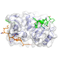
Native Data Set
Data DOI: 10.15785/SBGRID/402 | PDB ID 5EHM: RCSB PDBe | Publication DOI: 10.1016/j.neuron.2016.10.058 | Published: 13 Dec 2016
Mayer Laboratory, National Institutes of Health
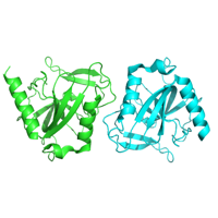
Native dataset
Data DOI: 10.15785/SBGRID/401 | PDB ID 5IUK: RCSB PDBe | Publication DOI: 10.7554/eLife.21422 | Published: 13 Dec 2016
Buschiazzo Laboratory, Institut Pasteur de Montevideo
Native dataset
Data DOI: 10.15785/SBGRID/400 | PDB ID 5IUN: RCSB PDBe | Publication DOI: 10.7554/eLife.21422 | Published: 13 Dec 2016
Buschiazzo Laboratory, Institut Pasteur de Montevideo
Native dataset
Data DOI: 10.15785/SBGRID/399 | PDB ID 5IUJ: RCSB PDBe | Publication DOI: 10.7554/eLife.21422 | Published: 13 Dec 2016
Buschiazzo Laboratory, Institut Pasteur de Montevideo
Native dates for Csm1 bound to Ulp2-Tof2 fusion
Data DOI: 10.15785/SBGRID/398 | PDB ID 53VN: RCSB PDBe | Published: 9 May 2017
Corbett Laboratory, University of California, San Diego
Native dataset of inorganic pyrophosphatase crystallised as a contaminant
Data DOI: 10.15785/SBGRID/393 | PDB ID 5H4F: RCSB PDBe | Published: 22 Nov 2016
Murthy Laboratory, Indian Institute of Science
x-ray images
Data DOI: 10.15785/SBGRID/392 | PDB ID 5FFO: RCSB PDBe | Published: 13 Dec 2016
Springer Laboratory, Children's Hospital Boston
X-ray diffraction
Data DOI: 10.15785/SBGRID/391 | PDB ID 5FFG: RCSB PDBe | Published: 13 Dec 2016
Springer Laboratory, Children's Hospital Boston
Native VapC4 dataset at 2.2A
Data DOI: 10.15785/SBGRID/390 | PDB ID 5H4H: RCSB PDBe | Published: 25 Nov 2016
Murthy Laboratory, Indian Institute of Science
Native VapC4 dataset at 1.77 A
Data DOI: 10.15785/SBGRID/389 | PDB ID 5H4G: RCSB PDBe | Published: 18 Nov 2016
Murthy Laboratory, Indian Institute of Science
Native dataset of a crystal which was obtained serendipitously. Now we know that the crystal corresponds to methylglyoxal synthase which was crystallised as a contaminant.
Data DOI: 10.15785/SBGRID/387 | PDB ID 5H3L: RCSB PDBe | Published: 15 Nov 2016
Murthy Laboratory, Indian Institute of Science
Native dataset, Co(II) and PPG bound
Data DOI: 10.15785/SBGRID/386 | PDB ID 5IAX: RCSB PDBe | Publication DOI: 10.1074/jbc.M116.725432 | Published: 11 Nov 2016
Dey Laboratory, University of Iowa
Native dataset, Co(II) bound and malonate
Data DOI: 10.15785/SBGRID/385 | PDB ID 5IAV: RCSB PDBe | Publication DOI: 10.1074/jbc.M116.725432 | Published: 11 Nov 2016
Dey Laboratory, University of Iowa
Native dataset, no metal bound
Data DOI: 10.15785/SBGRID/384 | PDB ID 5IAT: RCSB PDBe | Publication DOI: 10.1074/jbc.M116.725432 | Published: 11 Nov 2016
Dey Laboratory, University of Iowa
Lower resolution dataset of BaP4H with Cd(II) and AKG in C2221
Data DOI: 10.15785/SBGRID/383 | PDB ID 5HV4: RCSB PDBe | Publication DOI: 10.1107/S2059798316004198 | Published: 8 Nov 2016
Dey Laboratory, University of Iowa
High resolution dataset with Cd(II) in space group P3
Data DOI: 10.15785/SBGRID/382 | PDB ID 5HV0: RCSB PDBe | Publication DOI: 10.1107/S2059798316004198 | Published: 8 Nov 2016
Dey Laboratory, University of Iowa
Native dataset
Data DOI: 10.15785/SBGRID/381 | PDB ID 5T9T: RCSB PDBe | Publication DOI: 10.7554/eLife.20930 | Published: 1 Nov 2016
Shapiro Laboratory, Columbia University
Native dataset
Data DOI: 10.15785/SBGRID/380 | PDB ID 5SZR: RCSB PDBe | Publication DOI: 10.7554/eLife.20930 | Published: 1 Nov 2016
Shapiro Laboratory, Columbia University