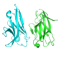
Native dataset
Data DOI: 10.15785/SBGRID/424 | PDB ID 5UTZ: RCSB PDBe | Published: 3 Jul 2018
Garcia Laboratory, Stanford University
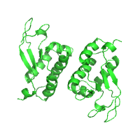
Native dataset
Data DOI: 10.15785/SBGRID/423 | PDB ID 5UN5: RCSB PDBe | Published: 7 Nov 2017
Garcia Laboratory, Stanford University
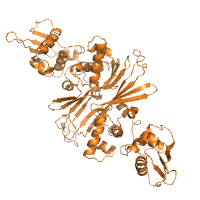
Native dataset
Data DOI: 10.15785/SBGRID/422 | PDB ID 5UN6: RCSB PDBe | Published: 7 Nov 2017
Garcia Laboratory, Stanford University
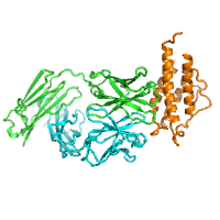
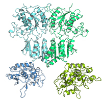
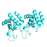
Native dataset
Data DOI: 10.15785/SBGRID/421 | PDB ID 5KVM: RCSB PDBe | Publication DOI: 10.1016/j.neuron.2016.08.022 | Published: 31 Jan 2017
Arac-Ozkan Laboratory, University of Chicago
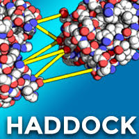
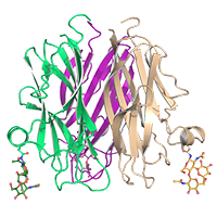
HADDOCK protein-peptide models gathered for the article: "A unified conformational selection and induced fit approach to protein-peptide docking." Trellet M, Melquiond ASJ, Bonvin AMJJ. Mar 2013. PLoS One 8(3) dx.doi.org/10.1371/journal.pone.0058769
Data DOI: 10.15785/SBGRID/420 | Publication DOI: 10.1371/journal.pone.0058769 | Published: 24 Jan 2017
Bonvin Laboratory, Utrecht University
Anisotropic data, with diffraction varying from 3.5 A to 7 A. First 400 images of a 600-image dataset, remainder of which had to be removed due to radiation damage. Crystal is twinned with a twin fraction of 0.49 and twinning law -h, -k, l. True space group is P3(2)21, but appears as P6(x)22 due to twinning.
Data DOI: 10.15785/SBGRID/419 | PDB ID 5L2E: RCSB PDBe | Publication DOI: 10.1016/j.str.2016.11.004 | Published: 20 Jan 2017
Ozkan Laboratory, University of Chicago
Rat Cerebellin-1, N-terminal domain degraded in the crystallization drops, resulting in the C1q-domain-only trimers.
Data DOI: 10.15785/SBGRID/418 | PDB ID 5KWR: RCSB PDBe | Publication DOI: 10.1016/j.str.2016.11.004 | Published: 17 Jan 2017
Ozkan Laboratory, University of Chicago
Single pulse (SASE) Free Electron Laser data collected from unexposed P6 sperm whale myoglobin crystals mounted in grids
Data DOI: 10.15785/SBGRID/417 | PDB ID 4PNJ: RCSB PDBe | Published: 17 Jan 2017
Macromolecular Crystallography Group Laboratory, Stanford Synchrotron Radiation Lightsource
Native dataset
Data DOI: 10.15785/SBGRID/416 | PDB ID 5T5W: RCSB PDBe | Published: 7 Nov 2017
Garcia Laboratory, Stanford University
native dataset
Data DOI: 10.15785/SBGRID/415 | PDB ID 5ELI: RCSB PDBe | Publication DOI: 10.7554/eLife.20391 | Published: 6 Jan 2017
Brett Laboratory, Washington U. School of Medicine
x-ray diffraction dataset, SeMet labeled protein, wavelength 0.9792
Data DOI: 10.15785/SBGRID/414 | PDB ID 5UBF: RCSB PDBe | Publication DOI: 10.1093/nar/gkw1288 | Published: 6 Jan 2017
Jeruzalmi Laboratory, City College of New York
x-ray diffraction dataset
Data DOI: 10.15785/SBGRID/413 | PDB ID 5UBD: RCSB PDBe | Publication DOI: 10.1093/nar/gkw1288 | Published: 6 Jan 2017
Jeruzalmi Laboratory, City College of New York
x-ray diffraction dataset
Data DOI: 10.15785/SBGRID/412 | PDB ID 5UBE: RCSB PDBe | Publication DOI: 10.1093/nar/gkw1288 | Published: 6 Jan 2017
Jeruzalmi Laboratory, City College of New York
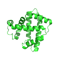
The crystal was obtained serendipitously
Data DOI: 10.15785/SBGRID/411 | PDB ID 5WQ5: RCSB PDBe | Published: 20 Dec 2016
Murthy Laboratory, Indian Institute of Science
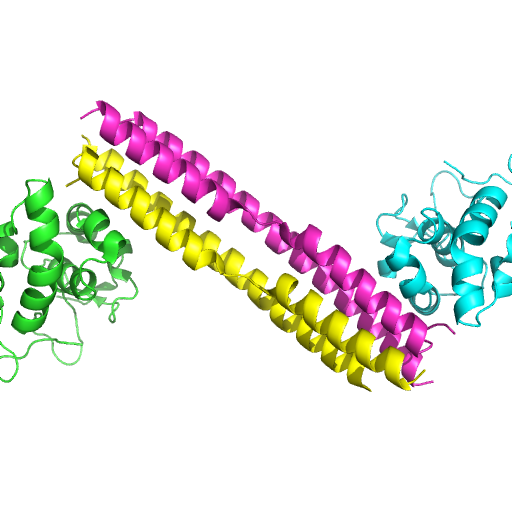
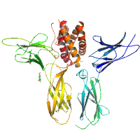
Native dataset for H. sapiens TRIP13 E253Q + ATP
Data DOI: 10.15785/SBGRID/410 | PDB ID 5VQA: RCSB PDBe | Published: 6 Jun 2017
Corbett Laboratory, University of California, San Diego
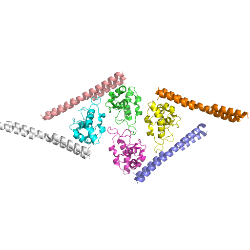
Native dataset for hTRIP13 E253Q Apo state
Data DOI: 10.15785/SBGRID/409 | PDB ID 5VQ9: RCSB PDBe | Published: 6 Jun 2017
Corbett Laboratory, University of California, San Diego
native dataset
Data DOI: 10.15785/SBGRID/408 | PDB ID 5IUL: RCSB PDBe | Publication DOI: 10.7554/eLife.21422 | Published: 13 Dec 2016
Buschiazzo Laboratory, Institut Pasteur de Montevideo
Native dataset
Data DOI: 10.15785/SBGRID/407 | PDB ID 5IUM: RCSB PDBe | Publication DOI: 10.7554/eLife.21422 | Published: 13 Dec 2016
Buschiazzo Laboratory, Institut Pasteur de Montevideo
Native dataset
Data DOI: 10.15785/SBGRID/406 | PDB ID 5ICT: RCSB PDBe | Publication DOI: 10.1016/j.neuron.2016.10.058 | Published: 13 Dec 2016
Mayer Laboratory, National Institutes of Health
Native dataset
Data DOI: 10.15785/SBGRID/405 | PDB ID 5DT6: RCSB PDBe | Publication DOI: 10.1016/j.neuron.2016.10.058 | Published: 13 Dec 2016
Mayer Laboratory, National Institutes of Health