HADDOCK models of mutant protein complexes gathered for the article: "C. Geng, A. Vangone, G.E. Folkers, L. Xue and Alexandre M.J.J. Bonvin, iSEE: Interface Structure, Evolution and Energy-based random forest predictor of binding affinity changes upon mutations. 2017. Submitted".
Data DOI: 10.15785/SBGRID/520 | Published: 5 Dec 2017
Bonvin Laboratory, Utrecht University
native dataset
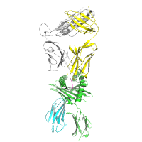
Data DOI: 10.15785/SBGRID/519 | PDB ID 6BJ3: RCSB PDBe | Published: 11 Jan 2019
Garcia Laboratory, Stanford University
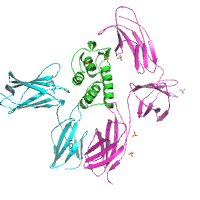
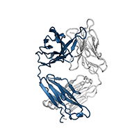
TCR589 in complex with HIV(Pol448-456)/HLA-B35
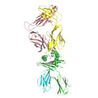
Data DOI: 10.15785/SBGRID/518 | PDB ID 6BJ2: RCSB PDBe | Published: 11 Jan 2019
Garcia Laboratory, Stanford University
native data
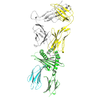
Data DOI: 10.15785/SBGRID/517 | PDB ID 6BJ8: RCSB PDBe | Published: 11 Jan 2019
Garcia Laboratory, Stanford University
native dataset
Data DOI: 10.15785/SBGRID/516 | PDB ID 5E4E: RCSB PDBe | Publication DOI: 10.1126/scisignal.aab2677 | Published: 7 Nov 2017
Garcia Laboratory, Stanford University
Native dataset
Data DOI: 10.15785/SBGRID/515 | PDB ID 6BGA: RCSB PDBe | Published: 11 Jan 2019
Garcia Laboratory, Stanford University
360 images
Data DOI: 10.15785/SBGRID/514 | PDB ID 6BHC: RCSB PDBe | Published: 12 Dec 2017
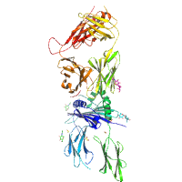
Boggon Laboratory, Yale University School of Medicine
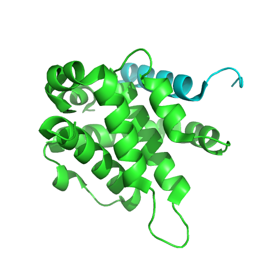
FPV039 in complex with Bik BH3 domain
Data DOI: 10.15785/SBGRID/513 | PDB ID 5TZP: RCSB PDBe | Published: 27 Oct 2017
Kvansakul Laboratory, La Trobe University
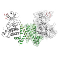
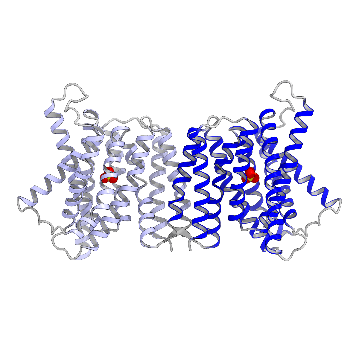
Triose-phosphate/phosphate translocator in complex with 3-phosphoglycerate
Data DOI: 10.15785/SBGRID/512 | PDB ID 5Y79: RCSB PDBe | Publication DOI: 10.1038/s41477-017-0022-8 | Published: 27 Oct 2017
Nureki Laboratory, The University of Tokyo
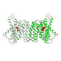
Triose-phosphate/phosphate translocator in complex with inorganic phosphate
Data DOI: 10.15785/SBGRID/511 | PDB ID 5Y78: RCSB PDBe | Publication DOI: 10.1038/s41477-017-0022-8 | Published: 27 Oct 2017
Nureki Laboratory, The University of Tokyo
X-ray diffraction images of the binding region of SK678.
Data DOI: 10.15785/SBGRID/510 | Published: 4 Jan 2022
Iverson Laboratory, Vanderbilt University
Diffraction data of the binding region of NCTC10712.
Data DOI: 10.15785/SBGRID/509 | Published: 4 Jan 2022
Iverson Laboratory, Vanderbilt University
Raw data for the binding region of SK150. Data were collected on a Bruker X8R at Vanderbilt University at room temperature.
Data DOI: 10.15785/SBGRID/508 | Published: 4 Jan 2022
Iverson Laboratory, Vanderbilt University
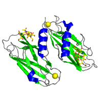
GspB siglec domain (with sialyl T antigen bound) in space group P 21 21 2
Data DOI: 10.15785/SBGRID/507 | PDB ID 5IUC: RCSB PDBe | Published: 20 Oct 2017
Iverson Laboratory, Vanderbilt University
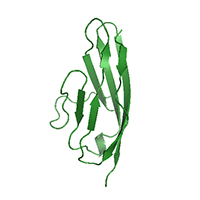
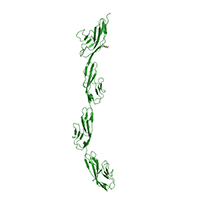
Robo1 Ig5 domain
Data DOI: 10.15785/SBGRID/505 | PDB ID 5O51: RCSB PDBe | Published: 23 Jan 2018
McCarthy Laboratory, EMBL - Grenoble
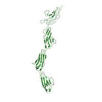
Robo1 Ig1-4 Crystal form 2
Data DOI: 10.15785/SBGRID/504 | PDB ID 5OPE: RCSB PDBe | Published: 23 Jan 2018
McCarthy Laboratory, EMBL - Grenoble
Robo1 Ig1-4 diffraction data collected on an ADSC Q315r detector
Data DOI: 10.15785/SBGRID/503 | PDB ID 5O5G: RCSB PDBe | Published: 23 Jan 2018
McCarthy Laboratory, EMBL - Grenoble
Dataset
Data DOI: 10.15785/SBGRID/500 | PDB ID 5TZP: RCSB PDBe | Publication DOI: 10.2210/pdb5tzp/pdb | Published: 3 Oct 2017
Kvansakul Laboratory, La Trobe University
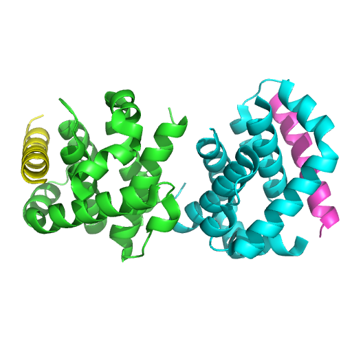
Dataset
Data DOI: 10.15785/SBGRID/499 | PDB ID 5TZQ: RCSB PDBe | Publication DOI: 10.2210/pdb5tzq/pdb | Published: 3 Oct 2017
Kvansakul Laboratory, La Trobe University
Native dataset
Data DOI: 10.15785/SBGRID/496 | PDB ID 5VZR: RCSB PDBe | Publication DOI: 10.1073/pnas.1707304114 | Published: 19 Sep 2017
McLellan Laboratory, University of Texas at Austin