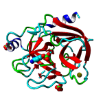
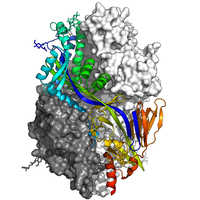
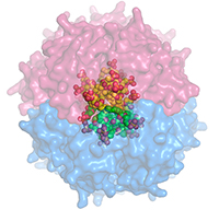
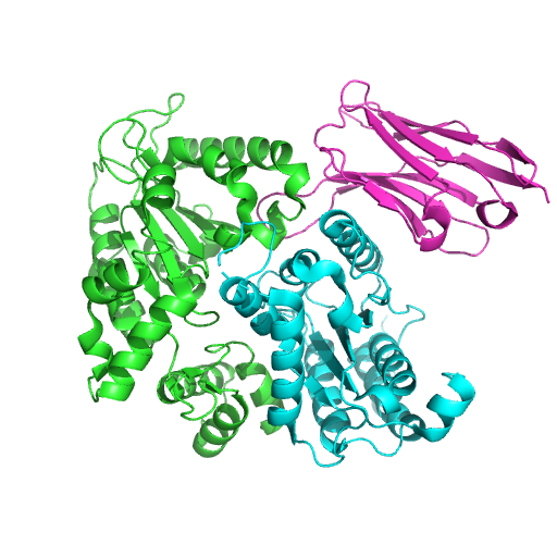
S-SAD Trypsin data collected on ID30B in two orientations (random and C* aligned)
Data DOI: 10.15785/SBGRID/541 | PDB ID 6fid: RCSB PDBe | Published: 23 Jan 2018
McCarthy Laboratory, EMBL - Grenoble
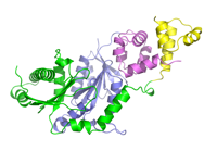
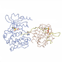
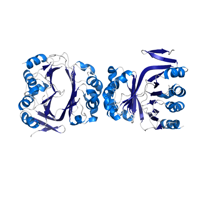
SeMet SAD dataset, C2 crystal form
Data DOI: 10.15785/SBGRID/540 | PDB ID 6BZG: RCSB PDBe | Published: 6 Feb 2018
Corbett Laboratory, University of California, San Diego
Native dataset, P212121 crystal form
Data DOI: 10.15785/SBGRID/539 | PDB ID 6BZG: RCSB PDBe | Published: 6 Feb 2018
Corbett Laboratory, University of California, San Diego
Native dataset, C2 crystal form
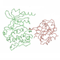
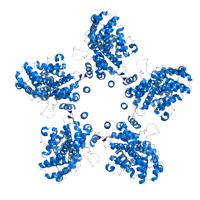
Data DOI: 10.15785/SBGRID/538 | PDB ID 6BZF: RCSB PDBe | Published: 6 Feb 2018
Corbett Laboratory, University of California, San Diego
Native
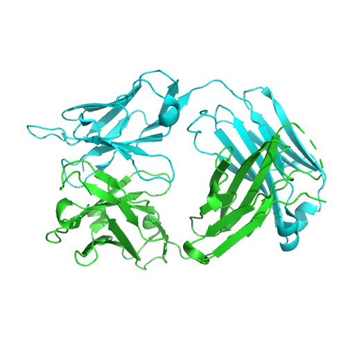
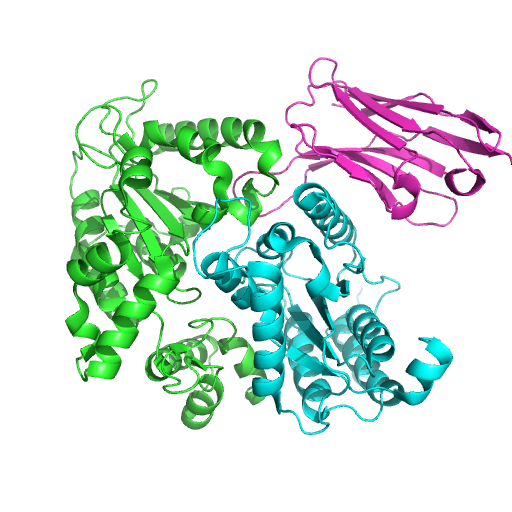
Data DOI: 10.15785/SBGRID/537 | PDB ID 5W24: RCSB PDBe | Publication DOI: 10.1038/s41467-017-01858-w | Published: 26 Dec 2017
McLellan Laboratory, University of Texas at Austin
Native dataset
Data DOI: 10.15785/SBGRID/536 | PDB ID 5W23: RCSB PDBe | Publication DOI: 10.1038/s41467-017-01858-w | Published: 26 Dec 2017
McLellan Laboratory, University of Texas at Austin
Native dataset
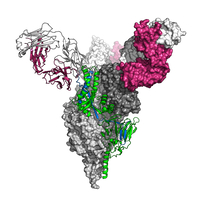
Data DOI: 10.15785/SBGRID/535 | PDB ID 5WB0: RCSB PDBe | Publication DOI: 10.1038/s41467-017-01708-9 | Published: 9 Jan 2018
McLellan Laboratory, University of Texas at Austin
Diffaction images
Data DOI: 10.15785/SBGRID/533 | PDB ID 5UPK: RCSB PDBe | Published: 22 Dec 2017
Boggon Laboratory, Yale University School of Medicine
Diffaction images
Data DOI: 10.15785/SBGRID/532 | PDB ID 5UPL: RCSB PDBe | Published: 22 Dec 2017
Boggon Laboratory, Yale University School of Medicine
SeMet data set
Data DOI: 10.15785/SBGRID/531 | PDB ID 6BTC: RCSB PDBe | Published: 17 Jul 2018
Rice Laboratory, University of Chicago
Native dataset
Data DOI: 10.15785/SBGRID/530 | PDB ID 6B55: RCSB PDBe | Published: 15 May 2018
Kvansakul Laboratory, La Trobe University
Native dataset
Data DOI: 10.15785/SBGRID/529 | PDB ID 5I2C: RCSB PDBe | Publication DOI: 10.1038/NATURE19079 | Published: 28 Nov 2017
Schwartz Laboratory, Massachusetts Institute of Technology
Native dataset
Data DOI: 10.15785/SBGRID/528 | PDB ID 5T0N: RCSB PDBe | Publication DOI: 10.1126/SCIENCE.AAD2087 | Published: 28 Nov 2017
Schwartz Laboratory, Massachusetts Institute of Technology
SeMet dataset
Data DOI: 10.15785/SBGRID/527 | PDB ID 5DJ4: RCSB PDBe | Publication DOI: 10.1126/SCIENCE.AAD2087 | Published: 28 Nov 2017
Schwartz Laboratory, Massachusetts Institute of Technology
Native dataset
Data DOI: 10.15785/SBGRID/526 | PDB ID 5DJ4: RCSB PDBe | Publication DOI: 10.1126/SCIENCE.AAD2087 | Published: 28 Nov 2017
Schwartz Laboratory, Massachusetts Institute of Technology
Native dataset
Data DOI: 10.15785/SBGRID/525 | PDB ID 5J1T: RCSB PDBe | Publication DOI: 10.7554/ELIFE.17983 | Published: 21 Nov 2017
Schwartz Laboratory, Massachusetts Institute of Technology
Native Data
Data DOI: 10.15785/SBGRID/524 | PDB ID 5J1S: RCSB PDBe | Publication DOI: 10.7554/eLife.17983 | Published: 21 Nov 2017
Schwartz Laboratory, Massachusetts Institute of Technology
X-ray diffraction data of crystals from Kluyveromyces lactis Ctf19-Mcm21; three data sets from native protein crystal
Data DOI: 10.15785/SBGRID/523 | PDB ID 3ZXU: RCSB PDBe | Publication DOI: 10.1038/embor.2012.1 | Published: 21 Nov 2017
Harrison Laboratory, Harvard Medical School
X-ray diffraction data of crystals from Kluyveromyces lactis central kinetochore proteins Ctf19-Mcm21; one anomalous data set from selenomethionine-derivatized protein crystal;
Data DOI: 10.15785/SBGRID/522 | PDB ID 3ZXU: RCSB PDBe | Publication DOI: 10.1038/embor.2012.1 | Published: 21 Nov 2017
Harrison Laboratory, Harvard Medical School
X-ray diffraction data of crystals of Kluyveromyces lactis central kinetochore proteins Ctf19-Mcm21 D-RWD domains bound with fragment of Kluyveromyces lactis central kinetochore protein Okp1
Data DOI: 10.15785/SBGRID/521 | PDB ID 5MU3: RCSB PDBe | Publication DOI: 10.15252/embj.201796636 | Published: 21 Nov 2017
Harrison Laboratory, Harvard Medical School