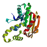
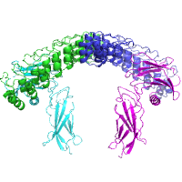
Native dataset
Data DOI: 10.15785/SBGRID/643 | PDB ID 6NRW: RCSB PDBe | Publication DOI: 10.7554/eLife.41028 | Published: 15 Feb 2019
Ozkan Laboratory, University of Chicago
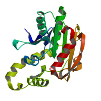
Native dataset
Data DOI: 10.15785/SBGRID/642 | PDB ID 6NRR: RCSB PDBe | Publication DOI: 10.7554/eLife.41028 | Published: 15 Feb 2019
Ozkan Laboratory, University of Chicago
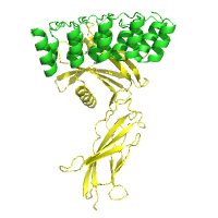
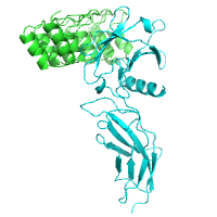
X-Ray Diffraction data from M. hassiacum GgH, source of 6Q5T structure
Data DOI: 10.15785/SBGRID/641 | PDB ID 6Q5T: RCSB PDBe | Published: 3 May 2019
Pereira Laboratory, IBMC/i3S, Universidade do Porto
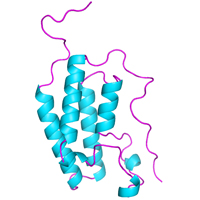
Complete data set from a single crystal for Lyn kinase SH3 domain
Data DOI: 10.15785/SBGRID/640 | PDB ID 6NMW: RCSB PDBe | Published: 18 Jan 2019
Iverson Laboratory, Vanderbilt University
Native Dataset
Data DOI: 10.15785/SBGRID/639 | PDB ID 6NE1: RCSB PDBe | Published: 24 May 2019
Garcia Laboratory, Stanford University
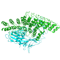
Merged datasets for structure solution by MR
Data DOI: 10.15785/SBGRID/638 | PDB ID 6NDZ: RCSB PDBe | Published: 24 May 2019
Garcia Laboratory, Stanford University
X-ray diffraction data for the RBCC motif of the tripartite motif (TRIM) of KAP1/TRIM28 collected at the zinc absorption peak wavelength
Data DOI: 10.15785/SBGRID/637 | PDB ID 6QAJ: RCSB PDBe | Published: 28 Jan 2020
Modis Laboratory, University of Cambridge
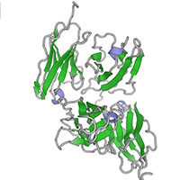
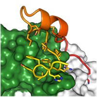
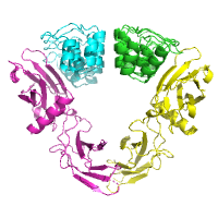
Complete data set from one single crystal used to solve the structure of the antibody-bound complex.
Data DOI: 10.15785/SBGRID/636 | PDB ID 6BLH: RCSB PDBe | Publication DOI: 10.1371/journal.ppat.1006935 | Published: 14 Dec 2018
McLellan Laboratory, University of Texas at Austin
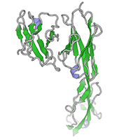
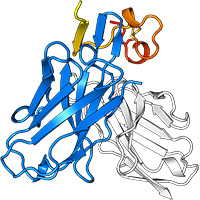
Complete data set from one crystal used to solve the structure of the G peptide-Fab complex.
Data DOI: 10.15785/SBGRID/635 | PDB ID 6BLI: RCSB PDBe | Publication DOI: 10.1371/journal.ppat.1006935 | Published: 14 Dec 2018
McLellan Laboratory, University of Texas at Austin
high resolution native data set
Data DOI: 10.15785/SBGRID/632 | PDB ID 6BA9: RCSB PDBe | Publication DOI: 10.1074/jbc.RA118.005752 | Published: 20 Nov 2018
Brett Laboratory, Washington U. School of Medicine
High resolution native data set
Data DOI: 10.15785/SBGRID/631 | PDB ID 6BA8: RCSB PDBe | Publication DOI: 10.1074/jbc.RA118.005752 | Published: 20 Nov 2018
Brett Laboratory, Washington U. School of Medicine
Native dataset
Data DOI: 10.15785/SBGRID/627 | PDB ID 5N15: RCSB PDBe | Publication DOI: 10.1038/ncomms15482 | Published: 30 Oct 2018
Petosa Laboratory, Institut de Biologie Structurale, Grenoble
Native Dataset
Data DOI: 10.15785/SBGRID/626 | PDB ID 6MOE: RCSB PDBe | Published: 24 May 2019
Garcia Laboratory, Stanford University
Native dataset
Data DOI: 10.15785/SBGRID/625 | PDB ID 6MOF: RCSB PDBe | Published: 24 May 2019
Garcia Laboratory, Stanford University
native dataset
Data DOI: 10.15785/SBGRID/624 | PDB ID 6MOG: RCSB PDBe | Published: 24 May 2019
Garcia Laboratory, Stanford University
Native Dataset
Data DOI: 10.15785/SBGRID/623 | PDB ID 6MOK: RCSB PDBe | Published: 24 May 2019
Garcia Laboratory, Stanford University
Native dataset
Data DOI: 10.15785/SBGRID/622 | PDB ID 6MOH: RCSB PDBe | Published: 24 May 2019
Garcia Laboratory, Stanford University
Native Dataset
Data DOI: 10.15785/SBGRID/621 | PDB ID 6MOI: RCSB PDBe | Published: 24 May 2019
Garcia Laboratory, Stanford University
native data set
Data DOI: 10.15785/SBGRID/620 | PDB ID 6MOJ: RCSB PDBe | Published: 24 May 2019
Garcia Laboratory, Stanford University
native dataset
Data DOI: 10.15785/SBGRID/619 | PDB ID 6MOL: RCSB PDBe | Published: 24 May 2019
Garcia Laboratory, Stanford University