Data DOI: 10.15785/SBGRID/834 | ID: 834
7MM1 Coordinates: Viewer, PDB (RCSB) (PDBe), MMDB
Hekstra Laboratory, Harvard University
Release Date: 18 May 2021
1. If this dataset is locally available, it should be accessable at /programs/datagrid/834
2. To download this dataset, please run the following command from your Terminal on a Linux or OS X workstation:
'rsync -av rsync://data.sbgrid.org/10.15785/SBGRID/834 .' (Harvard Medical School, USA)
Depending on your location, faster access may be available from a Tier 1 site closer to your location
'rsync -av rsync://sbgrid.icm.uu.se/10.15785/SBGRID/834 .' (Uppsala University, Sweden)
'rsync -av rsync://sbgrid.pasteur.edu.uy/10.15785/SBGRID/834 .' (Institut Pasteur de Montevideo, Uruguay)
'rsync -av rsync://sbgrid.ncpss.org/10.15785/SBGRID/834 .' (Shanghai Institutes for Biological Sciences, China)
3. After the transfer is completed, please issue the following command to verify data integrity:
'cd 834 ; shasum -c files.sha'
Storage requirements: 17G
Biological Sample:
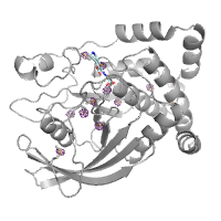
Protein tyrosine phosphatase 1B
Dataset Type:
X-Ray Diffraction
Subject Composition:
Protein
Collection Facility:
Beamline 24-ID-C, NE-CAT, APS, Argonne National Laboratory
Data Creation Date:
17 Jul 2019
Related Datasets:
None
Greisman, JB; Hekstra, D. 2021. "X-Ray Diffraction data for: Protein tyrosine phosphatase 1B. PDB Code 7MM1", SBGrid Data Bank, V1, https://doi.org/10.15785/SBGRID/834.
PTP1B in complex with TCS401 by Native S-SAD at Room Temperature
| Name | Additional Roles | Affiliation While Working on the Project |
|---|---|---|
| Jack B Greisman | Data Collector, Depositor | Harvard University |
| Doeke Hekstra | PI | Harvard University |
This data was collected with the Pilatus detector raised by two panels. This altered the beam center from that reported in the image headers and introduced a shadow on the detector. For data reduction, the beam center should be set to (290.5, 225.2), and the pixel mask provided in the "Processing Bundle" should be used. The experiment was conducted at 6.5 keV to improve the anomalous signal, but this introduced an additional harmonic in the incident X-ray beam. The resulting secondary diffraction pattern is only observed at low resolution, and was excluded from DIALS spot finding, indexing, and geometry refinement by setting d_max to 6 angstroms. The data was collected in two passes on the same crystal. Both passes were processed separately in DIALS and merged together in AIMLESS to yield the final merged intensities.
Version:
version unreported
Reprocessing failed.
Version:
version unreported
Reprocessing failed.
Version:
version unreported
Reprocessing failed.
License: CC0
Terms: Our Community Norms as well as good scientific practices expect that proper credit is given via citation. Please use the data citation, as generated by the SBGrid Data Bank.