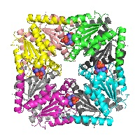
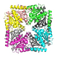
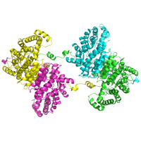
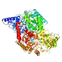
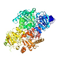
Structure of PurE (N5-carboxyaminoimidazole ribonucleotide mutase) H59N from the acidophilic bacterium Acetobacter aceti, bound to isocair
Data DOI: 10.15785/SBGRID/300 | PDB ID 2FWP: RCSB PDBe | Publication DOI: 10.1021/bi060465n | Published: 8 Jul 2016
Kappock Laboratory, Purdue University
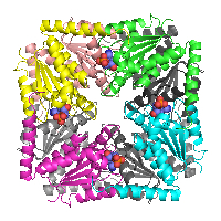
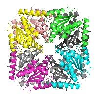
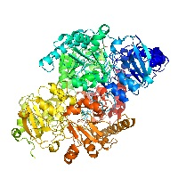
Structure of PurE (N5-carboxyaminoimidazole ribonucleotide mutase) from the acidophilic bacterium Acetobacter aceti, complexed with AIR (5-aminoimidazole ribonucleotide)
Data DOI: 10.15785/SBGRID/299 | PDB ID 2FWJ: RCSB PDBe | Publication DOI: 10.1021/bi060465n | Published: 31 May 2016
Kappock Laboratory, Purdue University
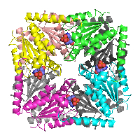
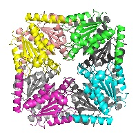
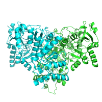
Structure of PurE (N5-carboxyaminoimidazole ribonucleotide mutase) H59D, from the acidophilic bacterium Acetobacter aceti, complexed with 5-aminoimidazole ribonucleotide (AIR)
Data DOI: 10.15785/SBGRID/298 | PDB ID 2FWI: RCSB PDBe | Publication DOI: 10.1021/bi060465n | Published: 31 May 2016
Kappock Laboratory, Purdue University
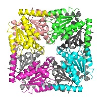
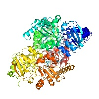
Structure of PurE (N5-carboxyaminoimidazole ribonucleotide mutase) H89F from the acidophilic bacterium Acetobacter aceti, at pH 8
Data DOI: 10.15785/SBGRID/297 | PDB ID 2FWB: RCSB PDBe | Publication DOI: 10.1021/bi060465n | Published: 31 May 2016
Kappock Laboratory, Purdue University
Structure of PurE (N5-carboxyaminoimidazole ribonucleotide mutase) H89N from the acidophilic bacterium Acetobacter aceti, at pH 7
Data DOI: 10.15785/SBGRID/296 | PDB ID 2FWA: RCSB PDBe | Publication DOI: 10.1021/bi060465n | Published: 31 May 2016
Kappock Laboratory, Purdue University
Structure of PurE (N5-carboxyaminoimidazole ribonucleotide mutase) H59F from the acidophilic bacterium Acetobacter aceti, at pH 8
Data DOI: 10.15785/SBGRID/295 | PDB ID 2FW9: RCSB PDBe | Publication DOI: 10.1021/bi060465n | Published: 31 May 2016
Kappock Laboratory, Purdue University
Structure of PurE (N5-carboxyaminoimidazole ribonucleotide mutase) H89G from the acidophilic bacterium Acetobacter aceti, at pH 8
Data DOI: 10.15785/SBGRID/294 | PDB ID 2FW8: RCSB PDBe | Publication DOI: 10.1021/bi060465n | Published: 31 May 2016
Kappock Laboratory, Purdue University
Structure of PurE (N5-carboxyaminoimidazole ribonucleotide mutase) H59N from the acidophilic bacterium Acetobacter aceti, at pH 8
Data DOI: 10.15785/SBGRID/293 | PDB ID 2FW7: RCSB PDBe | Publication DOI: 10.1021/bi060465n | Published: 31 May 2016
Kappock Laboratory, Purdue University
Structure of PurE (N5-carboxyaminoimidazole ribonucleotide mutase) mutant H59N from the acidophilic bacterium Acetobacter aceti, at pH 5.4
Data DOI: 10.15785/SBGRID/292 | PDB ID 2FW6: RCSB PDBe | Publication DOI: 10.1021/bi060465n | Published: 31 May 2016
Kappock Laboratory, Purdue University
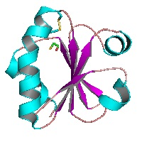
Crystal structure of thioredoxin from the acidophile Acetobacter aceti
Data DOI: 10.15785/SBGRID/291 | PDB ID 2I4A: RCSB PDBe | Publication DOI: 10.1110/ps.062519707 | Published: 8 Jul 2016
Kappock Laboratory, Purdue University
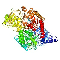
Structure of citrate synthase from the thermoacidophilic euryarchaeon Thermolasma acidophilum
Data DOI: 10.15785/SBGRID/283 | PDB ID 4YBO: RCSB PDBe | Publication DOI: 10.1107/S2053230X15015939 | Published: 27 May 2016
Kappock Laboratory, Purdue University
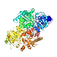
Succinyl-CoA:acetate CoA-transferase (AarCH6-N347A) in complex with CoA
Data DOI: 10.15785/SBGRID/282 | PDB ID 5DDK: RCSB PDBe | Publication DOI: 10.3389/fchem.2016.00023 | Published: 27 May 2016
Kappock Laboratory, Purdue University
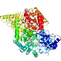
Succinyl-CoA:acetate CoA-transferase (AarC) in complex with CoA and citrate
Data DOI: 10.15785/SBGRID/281 | PDB ID 4EUD: RCSB PDBe | Publication DOI: 10.1021/bi300957f | Published: 27 May 2016
Kappock Laboratory, Purdue University
Succinyl-CoA:acetate CoA-transferase (AarCH6-E294A) in complex with dethiaacetyl-CoA
Data DOI: 10.15785/SBGRID/280 | PDB ID 4EUC: RCSB PDBe | Publication DOI: 10.1021/bi300957f | Published: 31 May 2016
Kappock Laboratory, Purdue University
Succinyl-CoA:acetate CoA-transferase (AarCH6-E294A) in complex with CoA
Data DOI: 10.15785/SBGRID/279 | PDB ID 4EUB: RCSB PDBe | Publication DOI: 10.1021/bi300957f | Published: 27 May 2016
Kappock Laboratory, Purdue University
Succinyl-CoA:acetate CoA-transferase (AarCH6-R228E) in complex with CoA and a covalent glutamyl-CoA thioester adduct
Data DOI: 10.15785/SBGRID/277 | PDB ID 4EU9: RCSB PDBe | Publication DOI: 10.1021/bi300957f | Published: 27 May 2016
Kappock Laboratory, Purdue University
Succinyl-CoA:acetate CoA-transferase (AarCH6-S71A) in complex with CoA
Data DOI: 10.15785/SBGRID/276 | PDB ID 4EU8: RCSB PDBe | Publication DOI: 10.1021/bi300957f | Published: 27 May 2016
Kappock Laboratory, Purdue University
Succinyl-CoA:acetate CoA-transferase (AarCH6) in complex with CoA and citrate
Data DOI: 10.15785/SBGRID/275 | PDB ID 4EU7: RCSB PDBe | Publication DOI: 10.1021/bi300957f | Published: 27 May 2016
Kappock Laboratory, Purdue University
Succinyl-CoA:acetate CoA-transferase (AarCH6) in complex with CoA, acetate, and covalent acetylglutamyl anhydride and glutamyl-CoA thioester adducts
Data DOI: 10.15785/SBGRID/274 | PDB ID 4EU6: RCSB PDBe | Publication DOI: 10.1021/bi300957f | Published: 27 May 2016
Kappock Laboratory, Purdue University
Succinyl-CoA:acetate CoA-transferase (AarCH6) in complex with CoA
Data DOI: 10.15785/SBGRID/273 | PDB ID 4EU5: RCSB PDBe | Publication DOI: 10.1021/bi300957f | Published: 27 May 2016
Kappock Laboratory, Purdue University