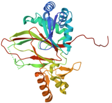
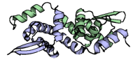
3 passes of 1440 raw diffraction images modeled by PDB ID 9B7C.
Data DOI: 10.15785/SBGRID/1276 | PDB ID 9B7C: RCSB PDBe | Publication DOI: 10.1126/sciadv.adj2921 | Published: 7 Jul 2026
Hekstra Laboratory, Harvard University
X-ray diffraction images for Glutarate hydroxylase (GlaH) from Escherichia coli
Data DOI: 10.15785/SBGRID/1271 | PDB ID 8CDF: RCSB PDBe | Published: 7 Jul 2026
Schubert Laboratory, University of Pretoria
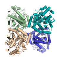
X-Ray Diffraction data from M. hassiacum GgH (Q434F mutant), source of 6G3N structure
Data DOI: 10.15785/SBGRID/1269 | PDB ID 6G3N: RCSB PDBe | Published: 8 May 2026
Pereira Laboratory, IBMC/i3S, Universidade do Porto
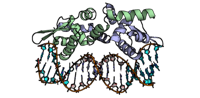
Crystal Structure of the Listeria monocytogenes CadC in complex with DNA
Data DOI: 10.15785/SBGRID/1261 | PDB ID 9T6T: RCSB PDBe | Published: 28 Apr 2026
Cabanes Laboratory, i3S
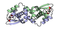
Crystal Structure of the Listeria monocytogenes CadC with Cadmium
Data DOI: 10.15785/SBGRID/1260 | PDB ID 9T6S: RCSB PDBe | Published: 28 Apr 2026
Cabanes Laboratory, i3S
Crystal Structure of the Listeria monocytogenes CadC
Data DOI: 10.15785/SBGRID/1259 | PDB ID 9T6R: RCSB PDBe | Published: 28 Apr 2026
Cabanes Laboratory, i3S
Native dataset. The crystal was macroscopically twinned.
Data DOI: 10.15785/SBGRID/1256 | PDB ID 8EVD: RCSB PDBe | Published: 10 Apr 2026
Madden Laboratory, Dartmouth Geisel School of Medicine
Native dataset
Data DOI: 10.15785/SBGRID/1241 | PDB ID 9Z7O: RCSB PDBe | Published: 26 May 2026
Corbett Laboratory, University of California, San Diego
Native dataset
Data DOI: 10.15785/SBGRID/1240 | PDB ID 9Z73: RCSB PDBe | Published: 26 May 2026
Corbett Laboratory, University of California, San Diego
Native dataset
Data DOI: 10.15785/SBGRID/1239 | PDB ID 9Z72: RCSB PDBe | Published: 26 May 2026
Corbett Laboratory, University of California, San Diego
Native dataset
Data DOI: 10.15785/SBGRID/1238 | PDB ID 9Z71: RCSB PDBe | Published: 26 May 2026
Corbett Laboratory, University of California, San Diego
Data collected at Vanadium peak wavelength
Data DOI: 10.15785/SBGRID/1235 | Published: 2 Dec 2025
Drennan Laboratory, Massachusetts Institute of Technology
Native dataset
Data DOI: 10.15785/SBGRID/1234 | PDB ID 9PCN: RCSB PDBe | Published: 2 Dec 2025
Drennan Laboratory, Massachusetts Institute of Technology
Native dataset
Data DOI: 10.15785/SBGRID/1233 | PDB ID 9PCM: RCSB PDBe | Published: 2 Dec 2025
Drennan Laboratory, Massachusetts Institute of Technology
Native dataset
Data DOI: 10.15785/SBGRID/1232 | PDB ID 9PCO: RCSB PDBe | Published: 2 Dec 2025
Drennan Laboratory, Massachusetts Institute of Technology
Native dataset
Data DOI: 10.15785/SBGRID/1231 | PDB ID 9PCL: RCSB PDBe | Published: 2 Dec 2025
Drennan Laboratory, Massachusetts Institute of Technology
Native dataset
Data DOI: 10.15785/SBGRID/1230 | PDB ID 9PC5: RCSB PDBe | Published: 2 Dec 2025
Drennan Laboratory, Massachusetts Institute of Technology
Native dataset
Data DOI: 10.15785/SBGRID/1229 | PDB ID 9PBX: RCSB PDBe | Published: 2 Dec 2025
Drennan Laboratory, Massachusetts Institute of Technology
Native dataset
Data DOI: 10.15785/SBGRID/1228 | PDB ID 9PBU: RCSB PDBe | Published: 2 Dec 2025
Drennan Laboratory, Massachusetts Institute of Technology
Native dataset
Data DOI: 10.15785/SBGRID/1227 | PDB ID 9PBN: RCSB PDBe | Published: 2 Dec 2025
Drennan Laboratory, Massachusetts Institute of Technology