native data, collected on four spots on the same crystal
Data DOI: 10.15785/SBGRID/181 | PDB ID 4FHM: RCSB PDBe | Publication DOI: 10.1073/pnas.1205151109 | Published: 6 Oct 2015
Schwartz Laboratory, Massachusetts Institute of Technology
Ta6Br12 derivative data
Data DOI: 10.15785/SBGRID/180 | PDB ID 4FHN: RCSB PDBe | Publication DOI: 10.1073/pnas.1205151109 | Published: 6 Oct 2015
Schwartz Laboratory, Massachusetts Institute of Technology
native data, collected in two separate spots
Data DOI: 10.15785/SBGRID/179 | PDB ID 4FHN: RCSB PDBe | Publication DOI: 10.1073/pnas.1205151109 | Published: 6 Oct 2015
Schwartz Laboratory, Massachusetts Institute of Technology
native data
Data DOI: 10.15785/SBGRID/178 | PDB ID 4FCC: RCSB PDBe | Publication DOI: 10.1073/pnas.1205151109 | Published: 6 Oct 2015
Schwartz Laboratory, Massachusetts Institute of Technology
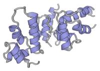
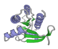
immature NTD domain (one extra residue at the N-terminus, before the mature Pro) / SAD dataset (quick iodide soak) collected at Cu Ka wavelength
Data DOI: 10.15785/SBGRID/177 | PDB ID 4PH3: RCSB PDBe | Publication DOI: 10.1126/science.aaa5182 | Published: 6 Oct 2015
Buschiazzo Laboratory, Institut Pasteur de Montevideo
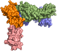
SeMet dataset, all proteins derivatized
Data DOI: 10.15785/SBGRID/176 | Publication DOI: 10.1038/nsmb.2998 | Published: 25 Sep 2015
Schwartz Laboratory, Massachusetts Institute of Technology
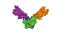
SeMet dataset, Nup85 derivatized only
Data DOI: 10.15785/SBGRID/175 | PDB ID 4YCZ: RCSB PDBe | Publication DOI: 10.1038/nsmb.2998 | Published: 25 Sep 2015
Schwartz Laboratory, Massachusetts Institute of Technology
Native dataset
Data DOI: 10.15785/SBGRID/174 | PDB ID 4ZI8: RCSB PDBe | Publication DOI: 10.1016/j.str.2015.09.005 | Published: 27 Oct 2015
Gaudet Laboratory, Harvard University
Native dataset
Data DOI: 10.15785/SBGRID/173 | PDB ID 4ZI9: RCSB PDBe | Publication DOI: 10.1016/j.str.2015.09.005 | Published: 27 Oct 2015
Gaudet Laboratory, Harvard University
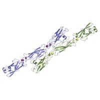
native data set
Data DOI: 10.15785/SBGRID/172 | PDB ID 4PH1: RCSB PDBe | Publication DOI: 10.1126/science.aaa5182 | Published: 8 Sep 2015
Buschiazzo Laboratory, Institut Pasteur de Montevideo
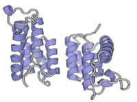
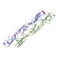
mature NTD domain / native data
Data DOI: 10.15785/SBGRID/171 | PDB ID 4PH2: RCSB PDBe | Publication DOI: 10.1126/science.aaa5182 | Published: 8 Sep 2015
Buschiazzo Laboratory, Institut Pasteur de Montevideo
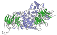
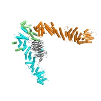
native (non-engineered) construct of full-length capsid
Data DOI: 10.15785/SBGRID/169 | PDB ID 4PH0: RCSB PDBe | Publication DOI: 10.1126/science.aaa5182 | Published: 8 Sep 2015
Buschiazzo Laboratory, Institut Pasteur de Montevideo
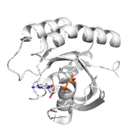
Crystal Structure of HIV-1 Reverse Transcriptase in Complex with 6-((4-((4-cyanophenyl)amino)-1,3,5-triazin-2-yl)amino)-5,7-dimethyl-2-naphthonitrile (JLJ639), a Non-nucleoside Inhibitor. 180 x 1 degree images collected for processing. Native data set collected on 24-ID-E at APS
Data DOI: 10.15785/SBGRID/168 | PDB ID 5C25: RCSB PDBe | Publication DOI: 10.1016/j.bmcl.2015.06.074 | Published: 8 Sep 2015
Anderson Laboratory, Yale University School of Medicine
SeMet dataset.
Data DOI: 10.15785/SBGRID/167 | PDB ID 4YD8: RCSB PDBe | Publication DOI: 10.1074/jbc.M115.649202 | Published: 18 Sep 2015
Schwartz Laboratory, Massachusetts Institute of Technology
SeMet dataset
Data DOI: 10.15785/SBGRID/166 | PDB ID 5DGK: RCSB PDBe | Published: 16 Aug 2016
Rice Laboratory, University of Chicago
native dataset
Data DOI: 10.15785/SBGRID/165 | PDB ID 4PGZ: RCSB PDBe | Publication DOI: 10.1016/j.molcel.2014.11.021 | Published: 6 Oct 2015
Schlessinger Laboratory, Yale University School of Medicine
native dataset
Data DOI: 10.15785/SBGRID/164 | PDB ID 4K94: RCSB PDBe | Publication DOI: 10.1073/pnas.1317118110 | Published: 6 Oct 2015
Schlessinger Laboratory, Yale University School of Medicine
Native data set
Data DOI: 10.15785/SBGRID/163 | PDB ID 4K9E: RCSB PDBe | Publication DOI: 10.1073/pnas.1317118110 | Published: 6 Oct 2015
Schlessinger Laboratory, Yale University School of Medicine
X-Ray diffraction data collected from a crystal of oncogenic mutant of human GTPase KRAS G12C bound to a novel GDP competitive covalent inhibitor, SML-8-73-1.
Data DOI: 10.15785/SBGRID/162 | PDB ID 4NMM: RCSB PDBe | Publication DOI: 10.1073/pnas.1404639111 | Published: 28 Aug 2015
Westover Laboratory, UT Southwestern Medical Center
X-Ray diffraction data collected from a crystal of oncogenic mutant of human GTPase KRAS Q61L.
Data DOI: 10.15785/SBGRID/161 | PDB ID 4WA7: RCSB PDBe | Publication DOI: 10.1158/1541-7786.MCR-15-0203 | Published: 28 Aug 2015
Westover Laboratory, UT Southwestern Medical Center