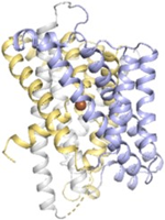
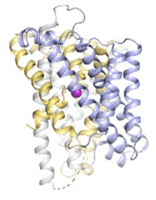
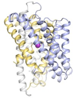
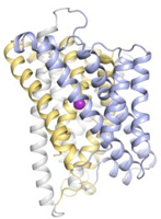
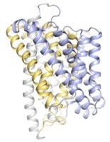
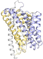
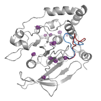
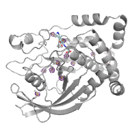
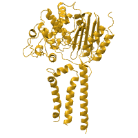
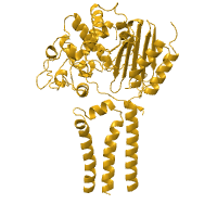
3 passes of 1440 raw diffraction images modeled by PDB ID 9B7C.
Data DOI: 10.15785/SBGRID/1276 | PDB ID 9B7C: RCSB PDBe | Publication DOI: 10.1126/sciadv.adj2921 | Published: 7 Jul 2026
Hekstra Laboratory, Harvard University
A series of 3049 pink-beam, polychromatic still images spaced one degree between adjacent frames. Metadata: Detector distance: 200 mm Beam center: (176.22 mm, 174.68 mm) Peak wavelength: 1.05 Angstroms Bandwidth: 5.5% (BioCARS) Unit cell constants: 79.405, 79.405, 37.837, 90.00, 90.00, 90.00 Space group: P43212
Data DOI: 10.15785/SBGRID/1118 | Published: 9 Jul 2024
Hekstra Laboratory, Harvard University
A series of 180 pink-beam, polychromatic still images spaced one degree between adjacent images. Metadata: Detector distance: 200 mm Beam center: (176.00 mm, 173.93 mm) Peak wavelength: 1.05 Angstroms Beam spectral bandwidth: 5.5% (BioCARS) Unit cell: 34.297, 45.552, 99.035, 90, 90, 90 Space group: P212121
Data DOI: 10.15785/SBGRID/1117 | Published: 9 Jul 2024
Hekstra Laboratory, Harvard University
Time-resolved data consisting of four passes of pink still images for PDZ2. Passes C, D, and E are spaced two degrees between adjacent images, with pass F spaced one degree between adjacent images. Start angles are 0, 92, 181, and 361.5 degrees for passes C, D, E, and F respectively. Time points are collected at 0, 50, 100, and 200 nanoseconds per image per pass. Metadata: Detector distance: 200 mm Beam center: (175.76 mm, 154.17 mm) Peak wavelength: 1.04 Angstroms Beam spectral bandwidth: 5.5% (BioCARS) Unit cell: 65.3, 39.45, 39.01, 90, 117.54, 90 Space group: C121 Applied voltage: 6.01 kV
Data DOI: 10.15785/SBGRID/1116 | PDB ID 5E11: RCSB PDBe | Publication DOI: https://doi.org/10.1038/nature20571 | Published: 9 Jul 2024
Hekstra Laboratory, Harvard University
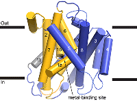
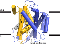
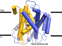
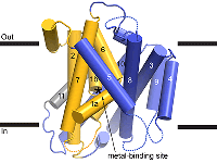
High resolution dataset collected from a single WT LCP crystal soaked in precipitant having cadmium.
Data DOI: 10.15785/SBGRID/969 | PDB ID 8E6M: RCSB PDBe | Published: 2 May 2023
Gaudet Laboratory, Harvard University
Datasets of Mn-bound D296A was collected from 3 LCP crystals. Image files that start C7run1_17 are from the first crystal, C7run2_19 are from the second crystal and C7run4_20 are from the third crystal.
Data DOI: 10.15785/SBGRID/968 | PDB ID 8E6L: RCSB PDBe | Published: 2 May 2023
Gaudet Laboratory, Harvard University
High resolution datasets of Mn-bound M230A mutant collected from 3 separate LCP crystals. Image files that start A131_run2 are from the first crystal, A132_run3 are from the second crystal and A133_run5 are from the third crystal.
Data DOI: 10.15785/SBGRID/967 | PDB ID 8E6I: RCSB PDBe | Published: 2 May 2023
Gaudet Laboratory, Harvard University
High resolution dataset of Mn-bound A47W mutant collected from a single LCP crystal
Data DOI: 10.15785/SBGRID/966 | PDB ID 8E6H: RCSB PDBe | Published: 2 May 2023
Gaudet Laboratory, Harvard University
High resolution dataset of a single LCP crystal of WT soaked in precipitant containing manganese
Data DOI: 10.15785/SBGRID/964 | PDB ID 8E60: RCSB PDBe | Published: 2 May 2023
Gaudet Laboratory, Harvard University
High resolution dataset of a single LCP crystal of WT mock-soaked in precipitant
Data DOI: 10.15785/SBGRID/963 | PDB ID 8E5V: RCSB PDBe | Published: 2 May 2023
Gaudet Laboratory, Harvard University
High resolution dataset of WT protein obtained from single LCP crystal
Data DOI: 10.15785/SBGRID/962 | PDB ID 8E5S: RCSB PDBe | Published: 2 May 2023
Gaudet Laboratory, Harvard University
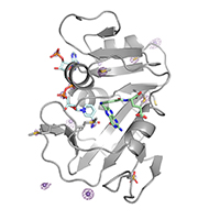
Apo PTP1B by Native S-SAD at Room Temperature
Data DOI: 10.15785/SBGRID/845 | PDB ID 7RIN: RCSB PDBe | Published: 31 Aug 2021
Hekstra Laboratory, Harvard University
PTP1B in complex with TCS401 by Native S-SAD at Room Temperature
Data DOI: 10.15785/SBGRID/834 | PDB ID 7MM1: RCSB PDBe | Published: 18 May 2021
Hekstra Laboratory, Harvard University
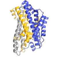
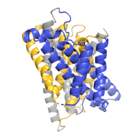
Data collected at the selenium edge of a single crystal of selenomethionine-substituted ClbP grown in a monoolein mesophase.
Data DOI: 10.15785/SBGRID/833 | PDB ID 7MDE: RCSB PDBe | Published: 4 Oct 2022
Gaudet Laboratory, Harvard University
Data collected from a single crystal of the wildtype ClbP peptidase covalently inhibited by a boronic acid and grown in a monopalmitolein mesophase.
Data DOI: 10.15785/SBGRID/832 | PDB ID 7MDC: RCSB PDBe | Published: 4 Oct 2022
Gaudet Laboratory, Harvard University
Bromine edge data collected from two crystals of the ClbP peptidase grown in presence of a substrate analog and in a monopalmitolein mesophase.
Data DOI: 10.15785/SBGRID/831 | PDB ID 7MDF: RCSB PDBe | Published: 4 Oct 2022
Gaudet Laboratory, Harvard University
E. coli DHFR by Native Mn,P,S-SAD at Room Temperature
Data DOI: 10.15785/SBGRID/821 | PDB ID 7LVC: RCSB PDBe | Published: 16 Mar 2021
Hekstra Laboratory, Harvard University
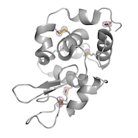
Hen Egg White Lysozyme by Native S-SAD at Room Temperature
Data DOI: 10.15785/SBGRID/816 | PDB ID 7L84: RCSB PDBe | Published: 22 Jan 2021
Hekstra Laboratory, Harvard University
native data set; best data set collected; contained multiple lattices
Data DOI: 10.15785/SBGRID/731 | PDB ID 6ORR: RCSB PDBe | Published: 6 Dec 2019
Gaudet Laboratory, Harvard University
39 wedges from LCP crystals collected with
Data DOI: 10.15785/SBGRID/576 | PDB ID 6D91: RCSB PDBe | Published: 5 Feb 2019
Gaudet Laboratory, Harvard University
Two wedges from separate LCP crystals were combined for a final data set that consisted of frames 1-400 of RG007_02_2 and frames 1-150 of RG007_03_1.
Data DOI: 10.15785/SBGRID/567 | PDB ID 6C3I: RCSB PDBe | Published: 5 Feb 2019
Gaudet Laboratory, Harvard University
Native dataset from 23 separate wedges from ~15 separate LCP crystals. Image files that start RG006_0101 are from the first wedge, RG006_0102 are from the second wedge etc. Only the frames from each wedge with usable diffraction data are included.
Data DOI: 10.15785/SBGRID/564 | PDB ID 6BU5: RCSB PDBe | Published: 5 Feb 2019
Gaudet Laboratory, Harvard University
Original images for 5UCG: Structure of the PP2C Phosphatase Domain and a Fragment of the Regulatory Domain of the Cell Fate Determinant SpoIIE from Bacillus Subtilis. From Bradshaw et al. eLife 2017;6:e26111.
Data DOI: 10.15785/SBGRID/469 | PDB ID 5UCG: RCSB PDBe | Publication DOI: 10.7554/eLife.26111 | Published: 21 Jul 2017
Gaudet Laboratory, Harvard University
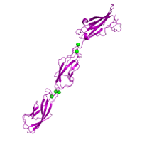
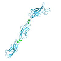
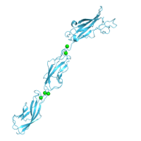
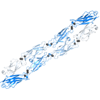
Native dataset used to determine the structure of Human Protocadherin-15 EC3-5 D414A Variant using molecular replacement.
Data DOI: 10.15785/SBGRID/363 | PDB ID 5T4N: RCSB PDBe | Published: 14 Feb 2017
Gaudet Laboratory, Harvard University
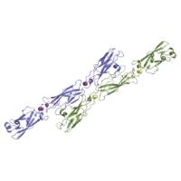
Native dataset used to determine low resolution structure of Mouse Protocadherin-15 EC4-5. The EC4-5 structure was then used as a molecular replacement search model to determine the structure of Human Protocadherin-15 EC3-5 (PDB Code: 5T4M)
Data DOI: 10.15785/SBGRID/362 | PDB ID 5T4M: RCSB PDBe | Published: 14 Feb 2017
Gaudet Laboratory, Harvard University
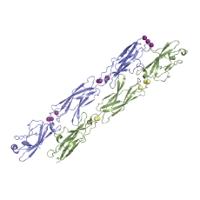
Native dataset of Human Protocadherin-15 EC3-5 used to determine structure using molecular replacement.
Data DOI: 10.15785/SBGRID/361 | PDB ID 5T4M: RCSB PDBe | Published: 14 Feb 2017
Gaudet Laboratory, Harvard University
Native dataset.
Data DOI: 10.15785/SBGRID/335 | Publication DOI: 10.1016/j.str.2016.09.017 | Published: 15 Nov 2016
Gaudet Laboratory, Harvard University
The second of two osmium containing data sets.
Data DOI: 10.15785/SBGRID/334 | PDB ID 5KTE: RCSB PDBe | Publication DOI: 10.1016/j.str.2016.09.017 | Published: 15 Nov 2016
Gaudet Laboratory, Harvard University
One of two Osmium containing data sets.
Data DOI: 10.15785/SBGRID/333 | PDB ID 5KTE: RCSB PDBe | Publication DOI: 10.1016/j.str.2016.09.017 | Published: 15 Nov 2016
Gaudet Laboratory, Harvard University
Native dataset.
Data DOI: 10.15785/SBGRID/332 | PDB ID 5KTE: RCSB PDBe | Publication DOI: 10.1016/j.str.2016.09.017 | Published: 15 Nov 2016
Gaudet Laboratory, Harvard University
Native data
Data DOI: 10.15785/SBGRID/325 | PDB ID 5K8R: RCSB PDBe | Published: 29 Jul 2016
Gaudet Laboratory, Harvard University
Native dataset
Data DOI: 10.15785/SBGRID/174 | PDB ID 4ZI8: RCSB PDBe | Publication DOI: 10.1016/j.str.2015.09.005 | Published: 27 Oct 2015
Gaudet Laboratory, Harvard University
Native dataset
Data DOI: 10.15785/SBGRID/173 | PDB ID 4ZI9: RCSB PDBe | Publication DOI: 10.1016/j.str.2015.09.005 | Published: 27 Oct 2015
Gaudet Laboratory, Harvard University
TRPV1-ARD native dataset
Data DOI: 10.15785/SBGRID/15 | PDB ID 2PNN: RCSB PDBe | Publication DOI: 10.1016/j.neuron.2007.05.027 | Published: 5 May 2015
Gaudet Laboratory, Harvard University
Seleno-methionine single wavelength anomalous diffraction dataset
Data DOI: 10.15785/SBGRID/14 | PDB ID 2NYJ: RCSB PDBe | Publication DOI: 10.1016/j.neuron.2007.05.027 | Published: 5 May 2015
Gaudet Laboratory, Harvard University