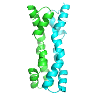
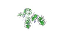
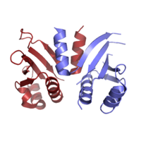
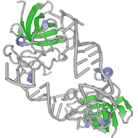
The set of all AlphaFold multimer (AF-M) v2.3 pairwise structure predictions accompanying the publication: Predictomes: A classifier-curated database of AlphaFold-modeled protein-protein interactions. This dataset includes prediction pairs used for training random forest classifiers including SPOC, pairs used for 30 ranking experiments, all pairs that belong to the genome maintenance matrix on predictomes.org, and three proteome wide in-silico interaction screens conducted with human DONSON, human STK19, and human USP37. All pairs were generated with ColabFold v1.5.2. All our predictions used AF-M multimer version 3 weights models 1, 2, and 4 with 3 recycles, templates enabled, 1 ensemble, no dropout, and no AMBER relaxation. The Multiple Sequence Alignments (MSAs) (unpaired + paired) supplied to AF-M were generated by the MMSeqs2 server using default settings. Sequences run were generally capped at 3,600 amino acids total to avoid memory exhaustion on GPUs.
Data DOI: 10.15785/SBGRID/1155 | Published: 4 Feb 2025
Walter Laboratory, Harvard Medical School
Native dataset, 0.979 angstroms
Data DOI: 10.15785/SBGRID/773 | PDB ID 6W1B: RCSB PDBe | Published: 17 Nov 2020
Rapoport Laboratory, Harvard Medical School
Native dataset #4 of 4 that were merged for structure solution
Data DOI: 10.15785/SBGRID/771 | PDB ID 6VZD: RCSB PDBe | Published: 17 Nov 2020
Rapoport Laboratory, Harvard Medical School
Native dataset #3 of 4 that were merged for structure solution
Data DOI: 10.15785/SBGRID/770 | PDB ID 6VZD: RCSB PDBe | Published: 17 Nov 2020
Rapoport Laboratory, Harvard Medical School
Native dataset #2 of 4 that were merged for structure solution
Data DOI: 10.15785/SBGRID/769 | PDB ID 6VZD: RCSB PDBe | Published: 17 Nov 2020
Rapoport Laboratory, Harvard Medical School
Native dataset #1 of 4 that were merged for structure solution
Data DOI: 10.15785/SBGRID/768 | PDB ID 6VZD: RCSB PDBe | Published: 17 Nov 2020
Rapoport Laboratory, Harvard Medical School
Native dataset, collected at 0.979 angstroms
Data DOI: 10.15785/SBGRID/767 | PDB ID 6VZE: RCSB PDBe | Published: 17 Nov 2020
Rapoport Laboratory, Harvard Medical School
Native dataset collected at 0.979 angstroms
Data DOI: 10.15785/SBGRID/766 | PDB ID 6VZ0: RCSB PDBe | Published: 17 Nov 2020
Rapoport Laboratory, Harvard Medical School
Sulfur-SAD dataset #7 (of 7 that were merged to create a dataset for structure solution)
Data DOI: 10.15785/SBGRID/763 | PDB ID 6VYN: RCSB PDBe | Published: 17 Nov 2020
Rapoport Laboratory, Harvard Medical School
Sulfur-SAD dataset #6 (of 7 that were merged to create a dataset for structure solution)
Data DOI: 10.15785/SBGRID/762 | PDB ID 6VYN: RCSB PDBe | Published: 17 Nov 2020
Rapoport Laboratory, Harvard Medical School
Sulfur-SAD dataset #5 (of 7 that were merged to create a dataset for structure solution)
Data DOI: 10.15785/SBGRID/761 | PDB ID 6VYN: RCSB PDBe | Published: 17 Nov 2020
Rapoport Laboratory, Harvard Medical School
Sulfur-SAD dataset #4 (of 7 that were merged to create a dataset for structure solution)
Data DOI: 10.15785/SBGRID/760 | PDB ID 6VYN: RCSB PDBe | Published: 17 Nov 2020
Rapoport Laboratory, Harvard Medical School
Sulfur-SAD dataset #3 (of 7 that were merged to create a dataset for structure solution)
Data DOI: 10.15785/SBGRID/759 | PDB ID 6VYN: RCSB PDBe | Published: 17 Nov 2020
Rapoport Laboratory, Harvard Medical School
Sulfur-SAD dataset #2 (of 7 that were merged to create a dataset for structure solution)
Data DOI: 10.15785/SBGRID/758 | PDB ID 6VYN: RCSB PDBe | Published: 17 Nov 2020
Rapoport Laboratory, Harvard Medical School
Sulfur-SAD dataset #1 (of 7 that were merged to create a dataset for structure solution)
Data DOI: 10.15785/SBGRID/757 | PDB ID 6VYN: RCSB PDBe | Published: 17 Nov 2020
Rapoport Laboratory, Harvard Medical School
Zn peak dataset
Data DOI: 10.15785/SBGRID/565 | PDB ID 6CDD: RCSB PDBe | Published: 19 Jun 2018
Rapoport Laboratory, Harvard Medical School
X-ray diffraction data of crystals from Kluyveromyces lactis Ctf19-Mcm21; three data sets from native protein crystal
Data DOI: 10.15785/SBGRID/523 | PDB ID 3ZXU: RCSB PDBe | Publication DOI: 10.1038/embor.2012.1 | Published: 21 Nov 2017
Harrison Laboratory, Harvard Medical School
X-ray diffraction data of crystals from Kluyveromyces lactis central kinetochore proteins Ctf19-Mcm21; one anomalous data set from selenomethionine-derivatized protein crystal;
Data DOI: 10.15785/SBGRID/522 | PDB ID 3ZXU: RCSB PDBe | Publication DOI: 10.1038/embor.2012.1 | Published: 21 Nov 2017
Harrison Laboratory, Harvard Medical School
X-ray diffraction data of crystals of Kluyveromyces lactis central kinetochore proteins Ctf19-Mcm21 D-RWD domains bound with fragment of Kluyveromyces lactis central kinetochore protein Okp1
Data DOI: 10.15785/SBGRID/521 | PDB ID 5MU3: RCSB PDBe | Publication DOI: 10.15252/embj.201796636 | Published: 21 Nov 2017
Harrison Laboratory, Harvard Medical School
single wavelength dataset of single crystal
Data DOI: 10.15785/SBGRID/265 | PDB ID 5IGQ: RCSB PDBe | Publication DOI: 10.1016/j.str.2016.03.002 | Published: 27 May 2016
Blacklow Laboratory, Harvard Medical School
single wavelength x ray data set of single crystal
Data DOI: 10.15785/SBGRID/264 | PDB ID 5IGO: RCSB PDBe | Publication DOI: 10.1016/j.str.2016.03.002 | Published: 27 May 2016
Blacklow Laboratory, Harvard Medical School
single wavelength dataset of single crystal
Data DOI: 10.15785/SBGRID/263 | PDB ID 5HQG: RCSB PDBe | Publication DOI: 10.1016/j.str.2016.03.002 | Published: 24 May 2016
Blacklow Laboratory, Harvard Medical School
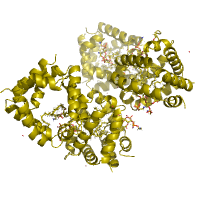
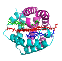
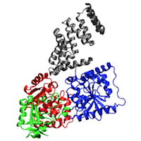
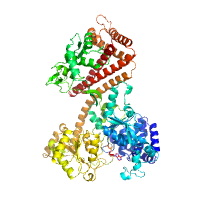
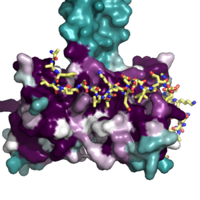
MD trajectory. The coordinates of the OGT–UDP–peptide complex (PDB 3PE4) were optimized in the Protein Preparation Wizard (Schrodinger 2009) where hydrogens were added; water molecules, UDP and peptide were stripped; and the structure was minimized using the OPLS2001 forcefield. The 1-μm simulation used the CHARM27 forcefield46, and the simple point charge model for water47. The CHARM27 forcefield was applied to the system using the VIPARR utility. The default Desmond relaxation was performed before simulation, and molecular dynamics were run at constant temperature (300 K) and pressure (1 bar). The simulation was performed by using the program Desmond, version 2.2.9.1.030 compiled by SBGrid on an optimized 64-node Linux-based InfiniBand cluster and took 75 days to complete. Molecular dynamics trajectories were processed and animated with VMD48.
Data DOI: 10.15785/SBGRID/190 | PDB ID 3PE4: RCSB PDBe | Publication DOI: 10.1038/nature09638 | Published: 3 Nov 2015
Sliz Laboratory, Harvard Medical School
Native dataset
Data DOI: 10.15785/SBGRID/184 | Publication DOI: 10.1021/jm501120z | Published: 23 Oct 2015
Blacklow Laboratory, Harvard Medical School
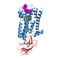
This dataset is compiled from 18 crystals of M2 receptor grown in the presence of the agonist iperoxo and the allosteric modulator LY2119620.
Data DOI: 10.15785/SBGRID/125 | PDB ID 4MQT: RCSB PDBe | Publication DOI: 10.1038/nature12735 | Published: 21 May 2015
Kruse Laboratory, Harvard Medical School
Native data
Data DOI: 10.15785/SBGRID/90 | PDB ID 4MLI: RCSB PDBe | Publication DOI: 10.1016/j.jmb.2013.10.021 | Published: 19 May 2015
Rapoport Laboratory, Harvard Medical School
Native dataset
Data DOI: 10.15785/SBGRID/80 | PDB ID 4YS0: RCSB PDBe | Publication DOI: 10.1016/j.jmb.2015.05.003 | Published: 19 May 2015
Rapoport Laboratory, Harvard Medical School
Native dataset, P21212 form
Data DOI: 10.15785/SBGRID/27 | PDB ID 3N4S: RCSB PDBe | Publication DOI: 10.1016/j.cell.2010.07.017 | Published: 6 May 2015
Harrison Laboratory, Harvard Medical School
Two-wedge SeMet peak wavelength dataset
Data DOI: 10.15785/SBGRID/26 | PDB ID 3N4R: RCSB PDBe | Publication DOI: 10.1016/j.cell.2010.07.017 | Published: 6 May 2015
Harrison Laboratory, Harvard Medical School
Native dataset
Data DOI: 10.15785/SBGRID/25 | PDB ID 3N7N: RCSB PDBe | Publication DOI: 10.1016/j.cell.2010.07.017 | Published: 6 May 2015
Harrison Laboratory, Harvard Medical School
Native dataset
Data DOI: 10.15785/SBGRID/24 | PDB ID 4EMC: RCSB PDBe | Publication DOI: 10.1016/j.celrep.2012.05.012 | Published: 6 May 2015
Harrison Laboratory, Harvard Medical School
Native dataset
Data DOI: 10.15785/SBGRID/4 | PDB ID 3TRZ: RCSB PDBe | Publication DOI: 10.1016/ j.cell.2011.10.020 | Published: 13 Apr 2015
Sliz Laboratory, Harvard Medical School
Zn-SAD dataset
Data DOI: 10.15785/SBGRID/3 | PDB ID 3TRZ: RCSB PDBe | Publication DOI: 10.1016/j.cell.2011.10.020 | Published: 13 Apr 2015
Sliz Laboratory, Harvard Medical School
native dataset
Data DOI: 10.15785/SBGRID/2 | PDB ID 3TS0: RCSB PDBe | Publication DOI: 10.1016/j.cell.2011.10.020 | Published: 10 Apr 2015
Sliz Laboratory, Harvard Medical School
Native Dataset
Data DOI: 10.15785/SBGRID/1 | PDB ID 3TS2: RCSB PDBe | Publication DOI: 10.1016/j.cell.2011.10.020 | Published: 10 Apr 2015
Sliz Laboratory, Harvard Medical School