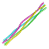
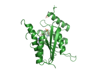
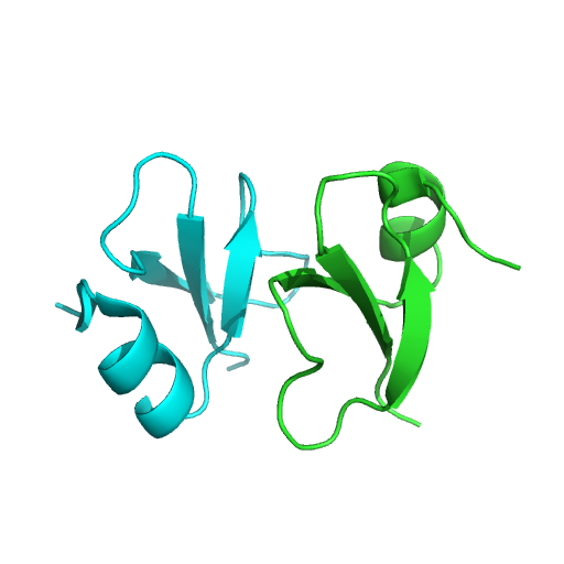
Selenomethionine SAD dataset for M. musculus SYCP3 105-248, P1 crystal form
Data DOI: 10.15785/SBGRID/584 | PDB ID 6DD9: RCSB PDBe | Published: 25 Jan 2019
Corbett Laboratory, University of California, San Diego
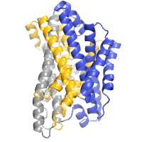
Selenomethionine SAD dataset for M. musculus SYCP3 105-248, P21 crystal form
Data DOI: 10.15785/SBGRID/583 | PDB ID 6DD8: RCSB PDBe | Published: 25 Jan 2019
Corbett Laboratory, University of California, San Diego
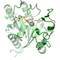
datasets merged from 2 crystals
Data DOI: 10.15785/SBGRID/582 | PDB ID 5WB2: RCSB PDBe | Published: 8 Jun 2018
Garcia Laboratory, Stanford University
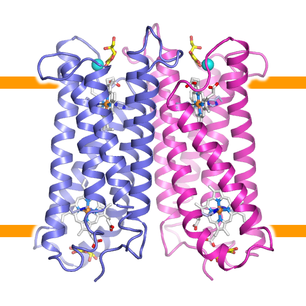
merged data from 2 crystals
Data DOI: 10.15785/SBGRID/581 | PDB ID 5WB1: RCSB PDBe | Published: 8 Jun 2018
Garcia Laboratory, Stanford University
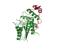
METTL16 MTase domain crystal from 2
Data DOI: 10.15785/SBGRID/579 | PDB ID 6GT5: RCSB PDBe | Published: 11 Sep 2018
McCarthy Laboratory, EMBL - Grenoble
METTL16 MTase domain crystal form 1
Data DOI: 10.15785/SBGRID/578 | PDB ID 6GFN: RCSB PDBe | Published: 11 Sep 2018
McCarthy Laboratory, EMBL - Grenoble
Diffraction data from crystal of del40 METTL16 MTase domain
Data DOI: 10.15785/SBGRID/577 | PDB ID 6GFK: RCSB PDBe | Published: 11 Sep 2018
McCarthy Laboratory, EMBL - Grenoble
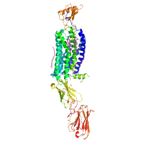
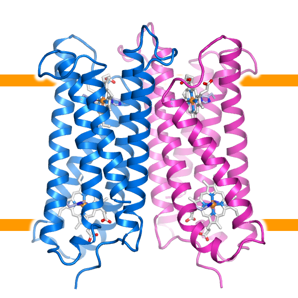
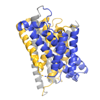
39 wedges from LCP crystals collected with
Data DOI: 10.15785/SBGRID/576 | PDB ID 6D91: RCSB PDBe | Published: 5 Feb 2019
Gaudet Laboratory, Harvard University
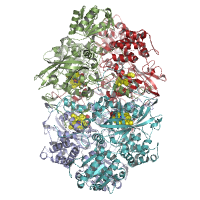
8 sweeps
Data DOI: 10.15785/SBGRID/575 | PDB ID 6D4G: RCSB PDBe | Published: 31 Aug 2018
Boggon Laboratory, Yale University School of Medicine
Small-wedge (5 degrees/crystal) data collected using 5x5 um beam
Data DOI: 10.15785/SBGRID/574 | PDB ID 5ZLG: RCSB PDBe | Published: 17 Aug 2018
Shiro Laboratory, University of Hyogo
Small-wedge (5 degrees/crystal) data collected using 5x5 um beam
Data DOI: 10.15785/SBGRID/573 | PDB ID 5ZLE: RCSB PDBe | Published: 17 Aug 2018
Shiro Laboratory, University of Hyogo
Native dataset
Data DOI: 10.15785/SBGRID/572 | PDB ID 6BY0: RCSB PDBe | Published: 30 Mar 2018
Buschiazzo Laboratory, Institut Pasteur de Montevideo
Native dataset
Data DOI: 10.15785/SBGRID/568 | PDB ID 6CS9: RCSB PDBe | Published: 27 Jul 2018
Kvansakul Laboratory, La Trobe University
Two wedges from separate LCP crystals were combined for a final data set that consisted of frames 1-400 of RG007_02_2 and frames 1-150 of RG007_03_1.
Data DOI: 10.15785/SBGRID/567 | PDB ID 6C3I: RCSB PDBe | Published: 5 Feb 2019
Gaudet Laboratory, Harvard University
HADDOCK refined models for the biological/crystallographic interfaces collected in the DC and MANY datasets
Data DOI: 10.15785/SBGRID/566 | Published: 6 Mar 2018
Bonvin Laboratory, Utrecht University
Zn peak dataset
Data DOI: 10.15785/SBGRID/565 | PDB ID 6CDD: RCSB PDBe | Published: 19 Jun 2018
Rapoport Laboratory, Harvard Medical School
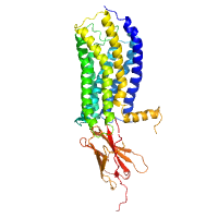
Native dataset from 23 separate wedges from ~15 separate LCP crystals. Image files that start RG006_0101 are from the first wedge, RG006_0102 are from the second wedge etc. Only the frames from each wedge with usable diffraction data are included.
Data DOI: 10.15785/SBGRID/564 | PDB ID 6BU5: RCSB PDBe | Published: 5 Feb 2019
Gaudet Laboratory, Harvard University
Native
Data DOI: 10.15785/SBGRID/563 | PDB ID 4Y5O: RCSB PDBe | Publication DOI: 10.1038/ncomms8937 | Published: 26 Jan 2018
Boggon Laboratory, Yale University School of Medicine
Native
Data DOI: 10.15785/SBGRID/562 | PDB ID 4WJ7: RCSB PDBe | Publication DOI: 10.1074/jbc.M114.616433 | Published: 26 Jan 2018
Boggon Laboratory, Yale University School of Medicine
Native
Data DOI: 10.15785/SBGRID/561 | PDB ID 4FQN: RCSB PDBe | Publication DOI: 10.1016/j.febslet.2012.12.011 | Published: 26 Jan 2018
Boggon Laboratory, Yale University School of Medicine