Data DOI: 10.15785/SBGRID/77 | ID: 77
Publication DOI: 10.1016/j.str.2013.06.003
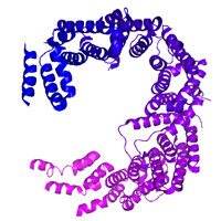
4BSN Coordinates: Viewer, PDB (RCSB) (PDBe), MMDB
Petosa Laboratory, Institut de Biologie Structurale, Grenoble
Release Date: 11 May 2015
1. If this dataset is locally available, it should be accessable at /programs/datagrid/77
2. To download this dataset, please run the following command from your Terminal on a Linux or OS X workstation:
'rsync -av rsync://data.sbgrid.org/10.15785/SBGRID/77 .' (Harvard Medical School, USA)
Depending on your location, faster access may be available from a Tier 1 site closer to your location
'rsync -av rsync://sbgrid.icm.uu.se/10.15785/SBGRID/77 .' (Uppsala University, Sweden)
'rsync -av rsync://sbgrid.pasteur.edu.uy/10.15785/SBGRID/77 .' (Institut Pasteur de Montevideo, Uruguay)
'rsync -av rsync://sbgrid.ncpss.org/10.15785/SBGRID/77 .' (Shanghai Institutes for Biological Sciences, China)
3. After the transfer is completed, please issue the following command to verify data integrity:
'cd 77 ; shasum -c files.sha'
Storage requirements: 16G
Biological Sample:
Human CRM1 (C-terminal truncation mutant)
Dataset Type:
X-Ray Diffraction
Subject Composition:
Protein
Collection Facility:
ESRF beamline ID23-2
Data Creation Date:
25 Apr 2009
Related Datasets:
None
Langer, K; Petosa, C. 2015. "X-Ray Diffraction data for: Human CRM1 (C-terminal truncation mutant). PDB Code 4BSN", SBGrid Data Bank, V1, https://doi.org/10.15785/SBGRID/77.
Native data for crystal variant 1: low resolution and high-resolution passes
| Name | Additional Roles | Affiliation While Working on the Project |
|---|---|---|
| Carlo Petosa | Data Collector, Depositor | Institut de Biologie Structurale, Grenoble, France |
| Karla Langer | Data Collector | Institut de Biologie Structurale, Grenoble, France |
| Carlo Petosa | PI | Institut de Biologie Structurale, Grenoble |
Crystals were highly sensitive to radiation damage and diffraction quality decayed rapidly. First, a low-resolution pass was collected in a single sweep of 180 degrees. (Files are named "lowres_1_xxx.mccd") Subsequently, several different volumes from the same crystal were exposed to higher dose over smaller oscillation ranges. (Files are named "highres_?_xxx.mccd", where ? is the run number.) Runs 8-11 were collected from a single crystal volume, whereas the other runs (1,2,4,5,6,7; there is no run 3) are from different volumes (i.e. a total of 7 crystal volumes were shot). The oscillation range for each image is given in the log file corresponding to each run. The structure factors deposited in the PDB were obtained from these image data by: i) integrating data from the different runs using different resolution limits (i.e., using lower resolution for those volumes that diffracted more poorly) and ii) integrating earlier images within each run using higher resolution limits than later images.
Version:
version unreported
Reprocessing failed.
Version:
version unreported
Reprocessing failed.
Version:
version unreported
Reprocessing failed.
License: CC0
Terms: Our Community Norms as well as good scientific practices expect that proper credit is given via citation. Please use the data citation, as generated by the SBGrid Data Bank.