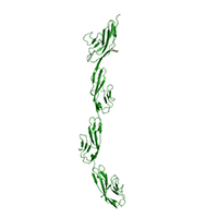
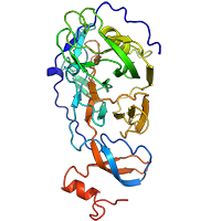
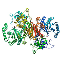
Robo1 Ig1-4 diffraction data collected on an ADSC Q315r detector
Data DOI: 10.15785/SBGRID/503 | PDB ID 5O5G: RCSB PDBe | Published: 23 Jan 2018
McCarthy Laboratory, EMBL - Grenoble
Dataset
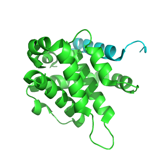
Data DOI: 10.15785/SBGRID/500 | PDB ID 5TZP: RCSB PDBe | Publication DOI: 10.2210/pdb5tzp/pdb | Published: 3 Oct 2017
Kvansakul Laboratory, La Trobe University
Dataset
Data DOI: 10.15785/SBGRID/499 | PDB ID 5TZQ: RCSB PDBe | Publication DOI: 10.2210/pdb5tzq/pdb | Published: 3 Oct 2017
Kvansakul Laboratory, La Trobe University
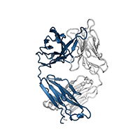
Native dataset
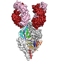
Data DOI: 10.15785/SBGRID/496 | PDB ID 5VZR: RCSB PDBe | Publication DOI: 10.1073/pnas.1707304114 | Published: 19 Sep 2017
McLellan Laboratory, University of Texas at Austin
Native dataset
Data DOI: 10.15785/SBGRID/495 | PDB ID 5VYH: RCSB PDBe | Publication DOI: 10.1073/pnas.1707304114 | Published: 19 Sep 2017
McLellan Laboratory, University of Texas at Austin
2 180 degree sweeps
Data DOI: 10.15785/SBGRID/494 | PDB ID 5VEF: RCSB PDBe | Published: 13 Oct 2017
Boggon Laboratory, Yale University School of Medicine
2 180 degree sweeps
Data DOI: 10.15785/SBGRID/493 | PDB ID 5VEE: RCSB PDBe | Published: 13 Oct 2017
Boggon Laboratory, Yale University School of Medicine
180 degrees
Data DOI: 10.15785/SBGRID/492 | PDB ID 5VED: RCSB PDBe | Published: 13 Oct 2017
Boggon Laboratory, Yale University School of Medicine
SeMet dataset
Data DOI: 10.15785/SBGRID/491 | PDB ID 5WHM: RCSB PDBe | Published: 29 Aug 2017
Crosson Laboratory, University of Chicago
Native dataset
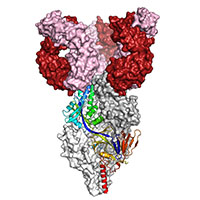
Data DOI: 10.15785/SBGRID/490 | PDB ID 5UDE: RCSB PDBe | Publication DOI: 10.1126/scitranslmed.aaj1928 | Published: 22 Aug 2017
McLellan Laboratory, University of Texas at Austin
Native dataset
Data DOI: 10.15785/SBGRID/489 | PDB ID 5UDD: RCSB PDBe | Publication DOI: 10.1126/scitranslmed.aaj1928 | Published: 22 Aug 2017
McLellan Laboratory, University of Texas at Austin
Native dataset
Data DOI: 10.15785/SBGRID/488 | PDB ID 5UDC: RCSB PDBe | Publication DOI: 10.1126/scitranslmed.aaj1928 | Published: 22 Aug 2017
McLellan Laboratory, University of Texas at Austin
X-Ray Diffraction data from human biliverdin IX beta reductase - erythrosine extra bluish complex, source of 5OOH structure - high resolution pass
Data DOI: 10.15785/SBGRID/487 | PDB ID 5OOH: RCSB PDBe | Published: 9 Mar 2018
Pereira Laboratory, IBMC/i3S, Universidade do Porto
X-Ray Diffraction data from human biliverdin IX beta reductase - erythrosine extra bluish complex, source of 5OOH structure - low resolution pass
Data DOI: 10.15785/SBGRID/486 | PDB ID 5OOH: RCSB PDBe | Published: 9 Mar 2018
Pereira Laboratory, IBMC/i3S, Universidade do Porto
X-Ray Diffraction data from human biliverdin IX beta reductase - phloxine B complex, source of 5OOG structure - high resolution pass
Data DOI: 10.15785/SBGRID/485 | PDB ID 5OOG: RCSB PDBe | Published: 9 Mar 2018
Pereira Laboratory, IBMC/i3S, Universidade do Porto
X-Ray Diffraction data from human biliverdin IX beta reductase - phloxine B complex, source of 5OOG structure - low resolution pass
Data DOI: 10.15785/SBGRID/484 | PDB ID 5OOG: RCSB PDBe | Published: 9 Mar 2018
Pereira Laboratory, IBMC/i3S, Universidade do Porto
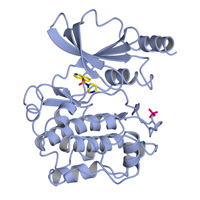
X-Ray Diffraction data from M. hassiacum GgH, source of 5OO2 structure
Data DOI: 10.15785/SBGRID/483 | PDB ID 5OO2: RCSB PDBe | Published: 3 May 2019
Pereira Laboratory, IBMC/i3S, Universidade do Porto
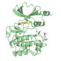
X-Ray Diffraction data from M. hassiacum GgH, source of 5ONZ structure
Data DOI: 10.15785/SBGRID/482 | PDB ID 5ONZ: RCSB PDBe | Published: 3 May 2019
Pereira Laboratory, IBMC/i3S, Universidade do Porto
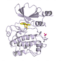
X-Ray Diffraction data from M. hassiacum GgH, source of 5ONT structure
Data DOI: 10.15785/SBGRID/481 | PDB ID 5ONT: RCSB PDBe | Published: 3 May 2019
Pereira Laboratory, IBMC/i3S, Universidade do Porto
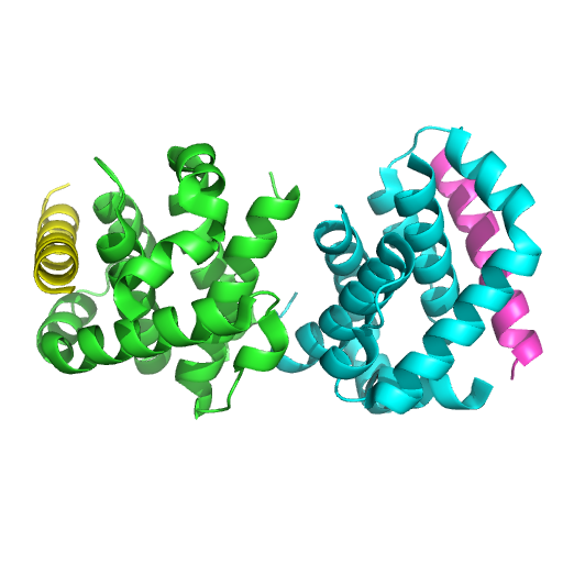
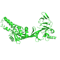
Native dataset
Data DOI: 10.15785/SBGRID/478 | PDB ID 5KWW: RCSB PDBe | Publication DOI: 10.1038/s41467-017-00170-x | Published: 28 Jul 2017
McLellan Laboratory, University of Texas at Austin