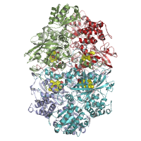
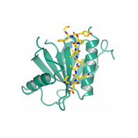
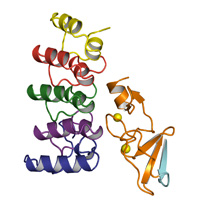
Native dataset
Data DOI: 10.15785/SBGRID/572 | PDB ID 6BY0: RCSB PDBe | Published: 30 Mar 2018
Buschiazzo Laboratory, Institut Pasteur de Montevideo
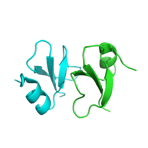
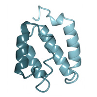
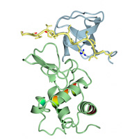
Native dataset
Data DOI: 10.15785/SBGRID/568 | PDB ID 6CS9: RCSB PDBe | Published: 27 Jul 2018
Kvansakul Laboratory, La Trobe University
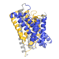
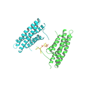
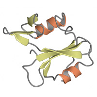
Two wedges from separate LCP crystals were combined for a final data set that consisted of frames 1-400 of RG007_02_2 and frames 1-150 of RG007_03_1.
Data DOI: 10.15785/SBGRID/567 | PDB ID 6C3I: RCSB PDBe | Published: 5 Feb 2019
Gaudet Laboratory, Harvard University
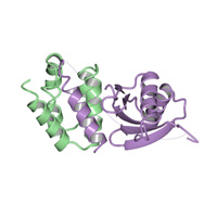
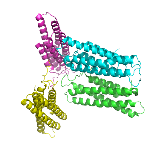
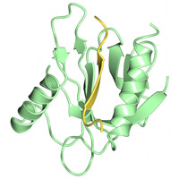
Zn peak dataset
Data DOI: 10.15785/SBGRID/565 | PDB ID 6CDD: RCSB PDBe | Published: 19 Jun 2018
Rapoport Laboratory, Harvard Medical School
Native dataset from 23 separate wedges from ~15 separate LCP crystals. Image files that start RG006_0101 are from the first wedge, RG006_0102 are from the second wedge etc. Only the frames from each wedge with usable diffraction data are included.
Data DOI: 10.15785/SBGRID/564 | PDB ID 6BU5: RCSB PDBe | Published: 5 Feb 2019
Gaudet Laboratory, Harvard University
Native
Data DOI: 10.15785/SBGRID/563 | PDB ID 4Y5O: RCSB PDBe | Publication DOI: 10.1038/ncomms8937 | Published: 26 Jan 2018
Boggon Laboratory, Yale University School of Medicine
Native
Data DOI: 10.15785/SBGRID/562 | PDB ID 4WJ7: RCSB PDBe | Publication DOI: 10.1074/jbc.M114.616433 | Published: 26 Jan 2018
Boggon Laboratory, Yale University School of Medicine
Native
Data DOI: 10.15785/SBGRID/561 | PDB ID 4FQN: RCSB PDBe | Publication DOI: 10.1016/j.febslet.2012.12.011 | Published: 26 Jan 2018
Boggon Laboratory, Yale University School of Medicine
Native Dataset
Data DOI: 10.15785/SBGRID/560 | PDB ID 4PR9: RCSB PDBe | Publication DOI: 10.1083/jcb.201404128 | Published: 26 Jan 2018
Tina Laboratory, The Scripps Research Institute
Native Dataset,
Data DOI: 10.15785/SBGRID/559 | PDB ID 5L0C: RCSB PDBe | Publication DOI: 10.1073/pnas.1600702113 | Published: 26 Jan 2018
Tina Laboratory, The Scripps Research Institute
Native
Data DOI: 10.15785/SBGRID/558 | PDB ID 4TVQ: RCSB PDBe | Publication DOI: 10.1083/jcb.201407129 | Published: 26 Jan 2018
Boggon Laboratory, Yale University School of Medicine
Native datasets
Data DOI: 10.15785/SBGRID/557 | PDB ID 6CDS: RCSB PDBe | Published: 28 Sep 2018
Tina Laboratory, The Scripps Research Institute
Native
Data DOI: 10.15785/SBGRID/556 | PDB ID 4DXA: RCSB PDBe | Publication DOI: 10.1074/jbc.M112.361295 | Published: 26 Jan 2018
Boggon Laboratory, Yale University School of Medicine
Native
Data DOI: 10.15785/SBGRID/555 | PDB ID 3IXE: RCSB PDBe | Publication DOI: 10.1016/j.jsb.2009.12.002 | Published: 26 Jan 2018
Boggon Laboratory, Yale University School of Medicine
Iodide soak
Data DOI: 10.15785/SBGRID/554 | PDB ID 3F6Q: RCSB PDBe | Publication DOI: 10.1073/pnas.0811415106 | Published: 26 Jan 2018
Boggon Laboratory, Yale University School of Medicine
Native
Data DOI: 10.15785/SBGRID/553 | PDB ID 3ULR: RCSB PDBe | Publication DOI: 10.1107/S1744309111056132 | Published: 26 Jan 2018
Boggon Laboratory, Yale University School of Medicine
Native
Data DOI: 10.15785/SBGRID/552 | PDB ID 4EIH: RCSB PDBe | Publication DOI: 10.1074/jbc.M114.556480 | Published: 26 Jan 2018
Boggon Laboratory, Yale University School of Medicine
Se SAD
Data DOI: 10.15785/SBGRID/550 | PDB ID 4DX9: RCSB PDBe | Publication DOI: 10.1016/j.molcel.2012.12.005 | Published: 23 Jan 2018
Boggon Laboratory, Yale University School of Medicine
Native
Data DOI: 10.15785/SBGRID/549 | PDB ID 4DX8: RCSB PDBe | Publication DOI: 10.1016/j.molcel.2012.12.005 | Published: 23 Jan 2018
Boggon Laboratory, Yale University School of Medicine
Native
Data DOI: 10.15785/SBGRID/548 | PDB ID 4JIF: RCSB PDBe | Published: 23 Jan 2018
Boggon Laboratory, Yale University School of Medicine