native data set
Data DOI: 10.15785/SBGRID/778 | PDB ID 5MGR: RCSB PDBe | Published: 13 May 2022
Dohnalek Laboratory, Institute of Biotechnology of the Czech Academy of Sciences
native data set
Data DOI: 10.15785/SBGRID/777 | PDB ID 5OHF: RCSB PDBe | Published: 21 Apr 2020
Dohnalek Laboratory, Institute of Biotechnology of the Czech Academy of Sciences
native data set
Data DOI: 10.15785/SBGRID/776 | PDB ID 5OHE: RCSB PDBe | Published: 21 Apr 2020
Dohnalek Laboratory, Institute of Biotechnology of the Czech Academy of Sciences
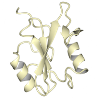
X-Ray Diffraction data from p120RasGAP C-terminal SH2 domain with p190RhoGAP pTyr-1087 peptide
Data DOI: 10.15785/SBGRID/775 | PDB ID 6WAY: RCSB PDBe | Published: 12 Jun 2020
Boggon Laboratory, Yale University School of Medicine
X-Ray Diffraction data from p120RasGAP C-terminal SH2 domain
Data DOI: 10.15785/SBGRID/774 | PDB ID 6WAX: RCSB PDBe | Published: 12 Jun 2020
Boggon Laboratory, Yale University School of Medicine
Native dataset, 0.979 angstroms
Data DOI: 10.15785/SBGRID/773 | PDB ID 6W1B: RCSB PDBe | Published: 17 Nov 2020
Rapoport Laboratory, Harvard Medical School
Native dataset #4 of 4 that were merged for structure solution
Data DOI: 10.15785/SBGRID/771 | PDB ID 6VZD: RCSB PDBe | Published: 17 Nov 2020
Rapoport Laboratory, Harvard Medical School
Native dataset #3 of 4 that were merged for structure solution
Data DOI: 10.15785/SBGRID/770 | PDB ID 6VZD: RCSB PDBe | Published: 17 Nov 2020
Rapoport Laboratory, Harvard Medical School
Native dataset #2 of 4 that were merged for structure solution
Data DOI: 10.15785/SBGRID/769 | PDB ID 6VZD: RCSB PDBe | Published: 17 Nov 2020
Rapoport Laboratory, Harvard Medical School
Native dataset #1 of 4 that were merged for structure solution
Data DOI: 10.15785/SBGRID/768 | PDB ID 6VZD: RCSB PDBe | Published: 17 Nov 2020
Rapoport Laboratory, Harvard Medical School
Native dataset, collected at 0.979 angstroms
Data DOI: 10.15785/SBGRID/767 | PDB ID 6VZE: RCSB PDBe | Published: 17 Nov 2020
Rapoport Laboratory, Harvard Medical School
Native dataset collected at 0.979 angstroms
Data DOI: 10.15785/SBGRID/766 | PDB ID 6VZ0: RCSB PDBe | Published: 17 Nov 2020
Rapoport Laboratory, Harvard Medical School
Sulfur-SAD dataset #7 (of 7 that were merged to create a dataset for structure solution)
Data DOI: 10.15785/SBGRID/763 | PDB ID 6VYN: RCSB PDBe | Published: 17 Nov 2020
Rapoport Laboratory, Harvard Medical School
Sulfur-SAD dataset #6 (of 7 that were merged to create a dataset for structure solution)
Data DOI: 10.15785/SBGRID/762 | PDB ID 6VYN: RCSB PDBe | Published: 17 Nov 2020
Rapoport Laboratory, Harvard Medical School
Sulfur-SAD dataset #5 (of 7 that were merged to create a dataset for structure solution)
Data DOI: 10.15785/SBGRID/761 | PDB ID 6VYN: RCSB PDBe | Published: 17 Nov 2020
Rapoport Laboratory, Harvard Medical School
Sulfur-SAD dataset #4 (of 7 that were merged to create a dataset for structure solution)
Data DOI: 10.15785/SBGRID/760 | PDB ID 6VYN: RCSB PDBe | Published: 17 Nov 2020
Rapoport Laboratory, Harvard Medical School
Sulfur-SAD dataset #3 (of 7 that were merged to create a dataset for structure solution)
Data DOI: 10.15785/SBGRID/759 | PDB ID 6VYN: RCSB PDBe | Published: 17 Nov 2020
Rapoport Laboratory, Harvard Medical School
Sulfur-SAD dataset #2 (of 7 that were merged to create a dataset for structure solution)
Data DOI: 10.15785/SBGRID/758 | PDB ID 6VYN: RCSB PDBe | Published: 17 Nov 2020
Rapoport Laboratory, Harvard Medical School
Sulfur-SAD dataset #1 (of 7 that were merged to create a dataset for structure solution)
Data DOI: 10.15785/SBGRID/757 | PDB ID 6VYN: RCSB PDBe | Published: 17 Nov 2020
Rapoport Laboratory, Harvard Medical School
native dataset
Data DOI: 10.15785/SBGRID/756 | PDB ID 6VS7: RCSB PDBe | Published: 1 Sep 2020
Iverson Laboratory, Vanderbilt University