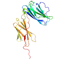
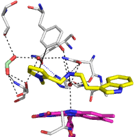
native
Data DOI: 10.15785/SBGRID/829 | PDB ID 6WDQ: RCSB PDBe | Publication DOI: https://doi.org/10.1016/j.cell.2021.01.018 | Published: 27 Apr 2021
Garcia Laboratory, Stanford University
SeMet dataset
Data DOI: 10.15785/SBGRID/828 | PDB ID 6WDP: RCSB PDBe | Publication DOI: https://doi.org/10.1016/j.cell.2021.01.018 | Published: 27 Apr 2021
Garcia Laboratory, Stanford University
Native dataset
Data DOI: 10.15785/SBGRID/827 | PDB ID 6WDP: RCSB PDBe | Publication DOI: 10.1016/j.cell.2021.01.018 | Published: 27 Apr 2021
Garcia Laboratory, Stanford University
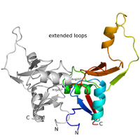
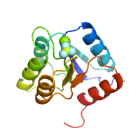
Zinc anomalous SAD dataset for V. polyspora Hop1 PHD-WHD domain
Data DOI: 10.15785/SBGRID/826 | PDB ID 7M0P: RCSB PDBe | Published: 4 Apr 2023
Corbett Laboratory, University of California, San Diego
This dataset comprises bound TCR-pMHC models for 44 TCR docking benchmark cases. These models were produced using four docking platforms - ClusPro, HADDOCK, LightDock and ZDOCK - as a comparative study of docking software performance in the context of TCR-pMHC modelling. Each docking case was provided to the software platforms along with varying levels of detail about the binding interface in the form of four docking scenarios, to assess how effectively each platform made use of this additional information to improve modeling accuracy. A manuscript reporting these results has been submitted for publication.
Data DOI: 10.15785/SBGRID/825 | Published: 21 May 2021
Chain Laboratory, University College London
native data set
Data DOI: 10.15785/SBGRID/824 | PDB ID 3M9Z: RCSB PDBe | Publication DOI: 10.1016/j.jsb.2011.05.001 | Published: 26 Mar 2021
Dohnalek Laboratory, Institute of Biotechnology of the Czech Academy of Sciences
E. coli DHFR by Native Mn,P,S-SAD at Room Temperature
Data DOI: 10.15785/SBGRID/821 | PDB ID 7LVC: RCSB PDBe | Published: 16 Mar 2021
Hekstra Laboratory, Harvard University
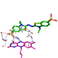
MicroED structure of a natural product VFAThiaGlu
Data DOI: 10.15785/SBGRID/820 | PDB ID 6PO6: RCSB PDBe | Publication DOI: 10.1126/science.aau6232 | Published: 29 Jan 2021
Gonen Laboratory, University of California, Los Angeles
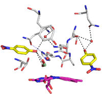
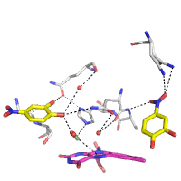
1.01 Å MicroED structure of GSNQNNF at 0.27 e- / A^2
Data DOI: 10.15785/SBGRID/819 | PDB ID 6CLC: RCSB PDBe | Publication DOI: 10.1016/j.str.2018.03.021 | Published: 29 Jan 2021
Gonen Laboratory, University of California, Los Angeles
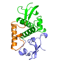
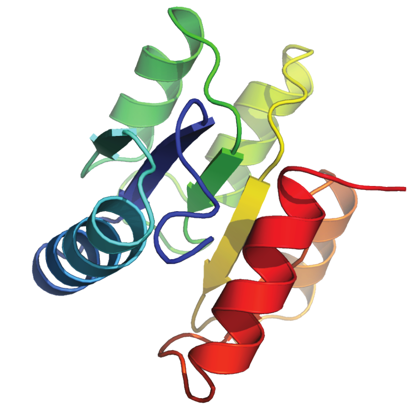
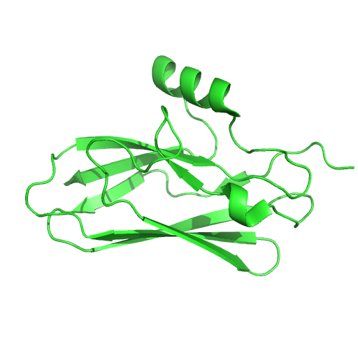
Receiver domain of RssB bound to beryllofluoride
Data DOI: 10.15785/SBGRID/818 | PDB ID 7LCM: RCSB PDBe | Published: 2 Apr 2021
Deaconescu Laboratory, Brown University
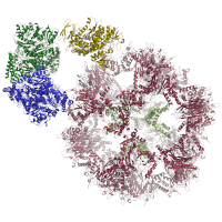
Structural models of the E1 and E2 proteins of Pyruvate Dehydrogenase Complex (PDHc), 2-Oxoglutarate Dehydrogenase (OGDHc), and Branched-Chain alpha-Keto Acid Dehydrogenase Complex (BCKDHc), E3BP of PDHc and E3, shared among all three complexes. In addition, a cif-file of E1, E2, E3BP, and E3 of PDHc modeled from cryoEM data is provided. Models were generated by homology modeling using MODELLER and refined using HADDOCK webserver.
Data DOI: 10.15785/SBGRID/817 | PDB ID 7BGJ: RCSB PDBe | Published: 26 Jan 2021
Kastritis Laboratory, Martin Luther University Halle-Wittenberg
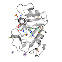
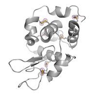
Hen Egg White Lysozyme by Native S-SAD at Room Temperature
Data DOI: 10.15785/SBGRID/816 | PDB ID 7L84: RCSB PDBe | Published: 22 Jan 2021
Hekstra Laboratory, Harvard University
Native dataset
Data DOI: 10.15785/SBGRID/815 | PDB ID 7L9C: RCSB PDBe | Published: 2 Apr 2021
Deaconescu Laboratory, Brown University
MicroED data of MyD88 TIR domain higher-order assembly microcrystals
Data DOI: 10.15785/SBGRID/814 | PDB ID 7BEQ: RCSB PDBe | Published: 12 Mar 2021
Zou Laboratory, Stockholm University
raw diffraction data
Data DOI: 10.15785/SBGRID/813 | PDB ID 7KMJ: RCSB PDBe | Published: 4 Jan 2022
Iverson Laboratory, Vanderbilt University
raw x-ray diffraction data
Data DOI: 10.15785/SBGRID/812 | PDB ID 6EF7: RCSB PDBe | Published: 3 Nov 2020
Iverson Laboratory, Vanderbilt University
native dataset
Data DOI: 10.15785/SBGRID/810 | PDB ID 7AA2: RCSB PDBe | Published: 18 May 2021
Dohnalek Laboratory, Institute of Biotechnology of the Czech Academy of Sciences
native dataset
Data DOI: 10.15785/SBGRID/809 | PDB ID 6ZE7: RCSB PDBe | Published: 18 May 2021
Dohnalek Laboratory, Institute of Biotechnology of the Czech Academy of Sciences
native dataset
Data DOI: 10.15785/SBGRID/808 | PDB ID 6ZE6: RCSB PDBe | Published: 18 May 2021
Dohnalek Laboratory, Institute of Biotechnology of the Czech Academy of Sciences
native dataset
Data DOI: 10.15785/SBGRID/807 | PDB ID 6ZE5: RCSB PDBe | Published: 18 May 2021
Dohnalek Laboratory, Institute of Biotechnology of the Czech Academy of Sciences