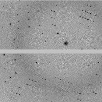
Sulfur-SAD dataset #2 (of 7 that were merged to create a dataset for structure solution)
Data DOI: 10.15785/SBGRID/758 | PDB ID 6VYN: RCSB PDBe | Published: 17 Nov 2020
Rapoport Laboratory, Harvard Medical School
Sulfur-SAD dataset #1 (of 7 that were merged to create a dataset for structure solution)
Data DOI: 10.15785/SBGRID/757 | PDB ID 6VYN: RCSB PDBe | Published: 17 Nov 2020
Rapoport Laboratory, Harvard Medical School
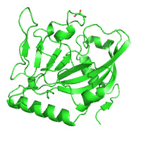
native dataset
Data DOI: 10.15785/SBGRID/756 | PDB ID 6VS7: RCSB PDBe | Published: 1 Sep 2020
Iverson Laboratory, Vanderbilt University
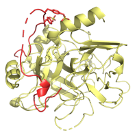
native dataset
Data DOI: 10.15785/SBGRID/755 | PDB ID 6VT2: RCSB PDBe | Published: 1 Sep 2020
Iverson Laboratory, Vanderbilt University
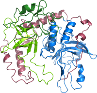
native dataset
Data DOI: 10.15785/SBGRID/754 | PDB ID 6VU6: RCSB PDBe | Published: 1 Sep 2020
Iverson Laboratory, Vanderbilt University
six data sets (six crystals) used for merging and molecular replacement (no experimental phasing)
Data DOI: 10.15785/SBGRID/753 | PDB ID 6YJP: RCSB PDBe | Published: 17 Jul 2020
Dohnalek Laboratory, Institute of Biotechnology of the Czech Academy of Sciences
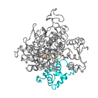
MicroED structure of a FIB-milled CypA Crystal; Tamir Gonen, Michael C Thompson, and James Fraser are co-PIs of this dataset.
Data DOI: 10.15785/SBGRID/752 | PDB ID 6U5G: RCSB PDBe | Published: 28 Jan 2020
Fraser Laboratory, University of California, San Francisco
This is the EXP data set referred to in the paper "Linking Crystallographic Model and Data Quality" by P. Andrew Karplus and Kay Diederichs, Science 336, 1030 (2012). The raw data for 3ELN were never deposited. The data deposited here are nicely fit by 3ELN, but they are about 15 times weaker than the original 3ELN data.
Data DOI: 10.15785/SBGRID/751 | PDB ID 3ELN: RCSB PDBe | Published: 28 Jan 2020
Diederichs Laboratory, University of Konstanz
Native dataset at 2.4 A resolution.
Data DOI: 10.15785/SBGRID/748 | PDB ID 6VAX: RCSB PDBe | Published: 1 Sep 2020
Iverson Laboratory, Vanderbilt University
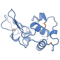
Hen lysozyme in triclinic space group at ambient temperature, diffuse scattering dataset
Data DOI: 10.15785/SBGRID/747 | PDB ID 6O2H: RCSB PDBe | Published: 28 Feb 2020
Ando Laboratory, Cornell University
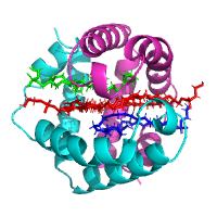
Simulated lysozyme data set - the diffraction images were generated by MLFSOM. The calculation of structure factors was based on the gadolinium derivative of tetragonal Hen Egg-White Lysozyme (PDB 1H87), all the alternative conformations of residues were removed.
Data DOI: 10.15785/SBGRID/746 | Published: 29 May 2020
Dohnalek Laboratory, Institute of Biotechnology of the Czech Academy of Sciences
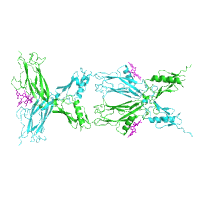
Native and Se-Met derivative data used for SAD structure determination.
Data DOI: 10.15785/SBGRID/742 | PDB ID 6TL1: RCSB PDBe | Published: 8 Sep 2020
Modis Laboratory, University of Cambridge
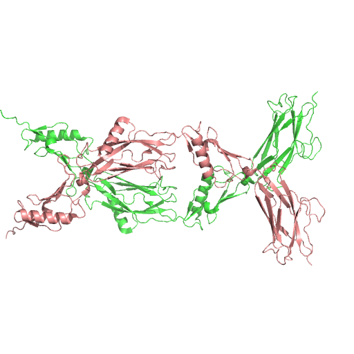
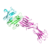
X-Ray Diffraction data from human thrombin:sulfated TTI complex, source of 6TKL structure
Data DOI: 10.15785/SBGRID/741 | PDB ID 6TKL: RCSB PDBe | Published: 27 Oct 2020
Pereira Laboratory, IBMC/i3S, Universidade do Porto
X-Ray Diffraction data from human thrombin:sulfated TTI complex, source of 6TKJ structure
Data DOI: 10.15785/SBGRID/740 | PDB ID 6TKJ: RCSB PDBe | Published: 27 Oct 2020
Pereira Laboratory, IBMC/i3S, Universidade do Porto
X-Ray Diffraction data from human thrombin:sulfated TTI complex, source of 6TKI structure
Data DOI: 10.15785/SBGRID/739 | PDB ID 6TKI: RCSB PDBe | Published: 27 Oct 2020
Pereira Laboratory, IBMC/i3S, Universidade do Porto
X-Ray Diffraction data from human thrombin:sulfated TTI complex, source of 6TKH structure
Data DOI: 10.15785/SBGRID/738 | PDB ID 6TKH: RCSB PDBe | Published: 27 Oct 2020
Pereira Laboratory, IBMC/i3S, Universidade do Porto
X-Ray Diffraction data from human thrombin:sulfated TTI complex, source of 6TKG structure
Data DOI: 10.15785/SBGRID/737 | PDB ID 6TKG: RCSB PDBe | Published: 27 Oct 2020
Pereira Laboratory, IBMC/i3S, Universidade do Porto
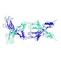
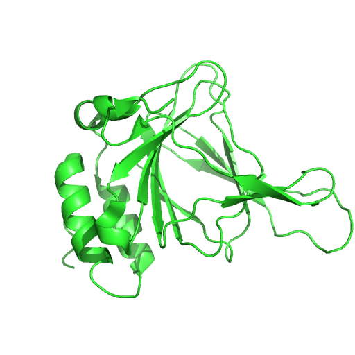
X-Ray Diffraction data from Photobacterium damselae subsp. piscicida NlpC/P60 endopeptidase, source of 6SQX structure
Data DOI: 10.15785/SBGRID/736 | PDB ID 6SQX: RCSB PDBe | Published: 14 Jul 2020
Pereira Laboratory, IBMC/i3S, Universidade do Porto
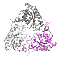
Native dataset for apo trimer form of Vibrio metoecus NucC
Data DOI: 10.15785/SBGRID/734 | PDB ID 6UXF: RCSB PDBe | Published: 20 Dec 2019
Corbett Laboratory, University of California, San Diego
Process to 1.45 A in XDS
Data DOI: 10.15785/SBGRID/732 | PDB ID 6Q1H: RCSB PDBe | Published: 20 Dec 2019
Corbett Laboratory, University of California, San Diego