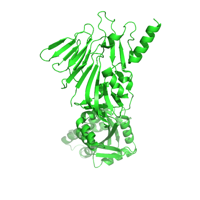
Native data set collected in two wedges
Data DOI: 10.15785/SBGRID/236 | PDB ID 5CNS: RCSB PDBe | Publication DOI: 10.7554/eLife.07141 | Published: 25 Mar 2016
Drennan Laboratory, Massachusetts Institute of Technology
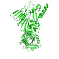
Best native dataset
Data DOI: 10.15785/SBGRID/235 | PDB ID 4MTK: RCSB PDBe | Published: 22 Apr 2016
Leiman Laboratory, École Polytechnique Fédérale de Lausanne
Native dataset
Data DOI: 10.15785/SBGRID/234 | PDB ID 5HAS: RCSB PDBe | Publication DOI: 10.7554/eLife.12411 | Published: 11 Mar 2016
Fromme Laboratory, Cornell University
SAD data of SeMet protein crystals. Data was collected in 10-image wedges using an inverse beam strategy.
Data DOI: 10.15785/SBGRID/233 | PDB ID 5HAS: RCSB PDBe | Publication DOI: 10.7554/eLife.12411 | Published: 11 Mar 2016
Fromme Laboratory, Cornell University
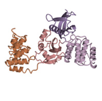
Data collected on Snf7 filament crystals after high-pressure freezing.
Data DOI: 10.15785/SBGRID/232 | PDB ID 5FD9: RCSB PDBe | Publication DOI: 10.7554/eLife.12548 | Published: 11 Mar 2016
Fromme Laboratory, Cornell University
Data collected on the Snf7 ESCRT-III filament at ambient pressure
Data DOI: 10.15785/SBGRID/231 | PDB ID 5FD7: RCSB PDBe | Publication DOI: 10.7554/eLife.12548 | Published: 11 Mar 2016
Fromme Laboratory, Cornell University
native dataset collected from single crystal of B. fragilis fragipain protease
Data DOI: 10.15785/SBGRID/225 | PDB ID 5DYN: RCSB PDBe | Publication DOI: 10.1038/nm.4077 | Published: 8 Mar 2016
Crosson Laboratory, University of Chicago
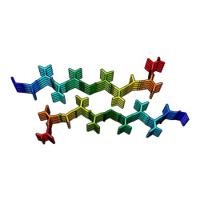
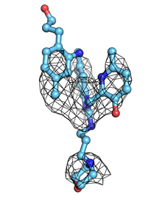
Structure of the amyloid forming segment, GAVVTGVTAVA, from the NAC domain of Parkinson's disease protein alpha-synuclein, residues 68-78, determined by electron diffraction.
Data DOI: 10.15785/SBGRID/223 | PDB ID 4RIL: RCSB PDBe | Publication DOI: 10.1038/nature15368 | Published: 4 Mar 2016
Gonen Laboratory, University of California, Los Angeles
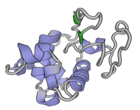
Lysozyme microcrystals
Data DOI: 10.15785/SBGRID/222 | PDB ID 3J6K: RCSB PDBe | Publication DOI: 10.1038/nmeth.3043 | Published: 4 Mar 2016
Gonen Laboratory, University of California, Los Angeles
THIS DATA IS ORIGINALLY USED IN XUE ET AL., BRIEFINGS IN BIOINFORMATICS, 2016: TEMPLATE-BASED PROTEIN-PROTEIN DOCKING EXPLOITING PAIRWISE INTERFACIAL RESIDUE RESTRAINTS by Li C Xue, Joao P.G.L.M. Rodrigues, Drena Dobbs, Vasant Honavar, Alexandre M.J.J. Bonvin
Data DOI: 10.15785/SBGRID/221 | Publication DOI: 10.1093/bib/bbw027 | Published: 9 Mar 2016
Bonvin Laboratory, Utrecht University
SeMet dataset.
Data DOI: 10.15785/SBGRID/220 | PDB ID 4YGA: RCSB PDBe | Publication DOI: 10.1073/pnas.1505914112 | Published: 15 Jan 2016
Schwartz Laboratory, Massachusetts Institute of Technology
Native data.
Data DOI: 10.15785/SBGRID/219 | PDB ID 4YGA: RCSB PDBe | Publication DOI: 10.1073/pnas.1505914112 | Published: 15 Jan 2016
Schwartz Laboratory, Massachusetts Institute of Technology
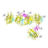
selenomethionyl neurexin 1alpha ectodomain
Data DOI: 10.15785/SBGRID/218 | PDB ID 3QCW: RCSB PDBe | Publication DOI: 10.1016/j.str.2011.03.012 | Published: 12 Jan 2016
Rudenko Laboratory, University of Texas Medical Branch
Native dataset
Data DOI: 10.15785/SBGRID/217 | PDB ID 5EA8: RCSB PDBe | Publication DOI: 10.1038/nchembio.1982 | Published: 8 Jan 2016
McLellan Laboratory, University of Texas at Austin
Native dataset
Data DOI: 10.15785/SBGRID/216 | PDB ID 5EA7: RCSB PDBe | Publication DOI: 10.1038/nchembio.1982 | Published: 8 Jan 2016
McLellan Laboratory, University of Texas at Austin
Native dataset
Data DOI: 10.15785/SBGRID/215 | PDB ID 5EA6: RCSB PDBe | Publication DOI: 10.1038/nchembio.1982 | Published: 8 Jan 2016
McLellan Laboratory, University of Texas at Austin
Native dataset
Data DOI: 10.15785/SBGRID/214 | PDB ID 5EA5: RCSB PDBe | Publication DOI: 10.1038/nchembio.1982 | Published: 8 Jan 2016
McLellan Laboratory, University of Texas at Austin
Native dataset
Data DOI: 10.15785/SBGRID/213 | PDB ID 5EA4: RCSB PDBe | Publication DOI: 10.1038/nchembio.1982 | Published: 8 Jan 2016
McLellan Laboratory, University of Texas at Austin
Native dataset
Data DOI: 10.15785/SBGRID/212 | PDB ID 5EA3: RCSB PDBe | Publication DOI: 10.1038/nchembio.1982 | Published: 8 Jan 2016
McLellan Laboratory, University of Texas at Austin
180°, 1° oscillations.
Data DOI: 10.15785/SBGRID/211 | PDB ID 5D68: RCSB PDBe | Publication DOI: 10.1016/j.jsb.2015.10.006 | Published: 5 Jan 2016
Boggon Laboratory, Yale University School of Medicine