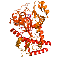
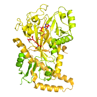
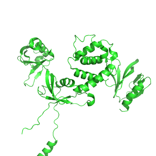
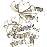
Engineered mouse Tubulin Tyrosine Ligase Like Enzyme 6 in complex with Initiation analog
Data DOI: 10.15785/SBGRID/803 | PDB ID 6VZR: RCSB PDBe | Publication DOI: 10.1038/s41594-020-0462-0 | Published: 14 Aug 2020
Roll-Mecak Laboratory, National Institutes of Health
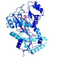
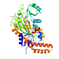
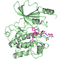
engineered mouse Tubulin Tyrosine Ligase Like Enzyme 6 in complex with gamma-elongation analog
Data DOI: 10.15785/SBGRID/802 | PDB ID 6VZS: RCSB PDBe | Publication DOI: 10.1038/s41594-020-0462-0 | Published: 14 Aug 2020
Roll-Mecak Laboratory, National Institutes of Health
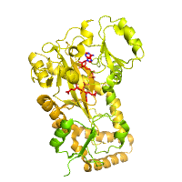
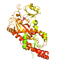
Engineered mouse Tubulin Tyrosine Ligase Like Enzyme 6 in complex with alpha-elongation analog
Data DOI: 10.15785/SBGRID/801 | PDB ID 6VZQ: RCSB PDBe | Publication DOI: 10.1038/s41594-020-0462-0 | Published: 14 Aug 2020
Roll-Mecak Laboratory, National Institutes of Health
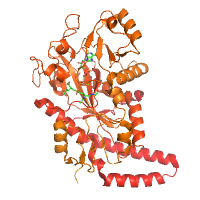
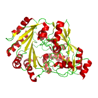
mouse Tubulin Tyrosine Ligase Like Enzyme 6 in complex with the Initiation analog
Data DOI: 10.15785/SBGRID/800 | PDB ID 6VZW: RCSB PDBe | Published: 14 Aug 2020
Roll-Mecak Laboratory, National Institutes of Health
mouse Tubulin Tyrosine Ligase Like Enzyme 6 in complex with gamma-elongation analog
Data DOI: 10.15785/SBGRID/799 | PDB ID 6VZV: RCSB PDBe | Published: 14 Aug 2020
Roll-Mecak Laboratory, National Institutes of Health
mouse Tubulin Tyrosine Ligase Like Enzyme 6 in complex with alpha-elongation analog
Data DOI: 10.15785/SBGRID/798 | PDB ID 6VZU: RCSB PDBe | Published: 14 Aug 2020
Roll-Mecak Laboratory, National Institutes of Health
mouse Tubulin Tyrosine Ligase Like Enzyme 6 in complex with ATP
Data DOI: 10.15785/SBGRID/797 | PDB ID 6VZT: RCSB PDBe | Published: 14 Aug 2020
Roll-Mecak Laboratory, National Institutes of Health
native data set
Data DOI: 10.15785/SBGRID/796 | PDB ID 3RJ8: RCSB PDBe | Published: 10 Jul 2020
Dohnalek Laboratory, Institute of Biotechnology of the Czech Academy of Sciences
Native Data Set
Data DOI: 10.15785/SBGRID/795 | PDB ID 6U4K: RCSB PDBe | Publication DOI: 10.1074/jbc.RA119.010789 | Published: 10 Jul 2020
Tina Laboratory, The Scripps Research Institute
Native data set
Data DOI: 10.15785/SBGRID/794 | PDB ID 6XF9: RCSB PDBe | Published: 19 Feb 2021
Glaunsinger Laboratory, University of California, Berkeley
Microcrystal electron diffraction data of HCA II
Data DOI: 10.15785/SBGRID/793 | PDB ID 6YMB: RCSB PDBe | Published: 4 Aug 2020
Zou Laboratory, Stockholm University
Microcrystal electron diffraction data of HCA II:AZM
Data DOI: 10.15785/SBGRID/792 | PDB ID 6YMA: RCSB PDBe | Published: 4 Aug 2020
Zou Laboratory, Stockholm University
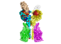
242 structural models of human ACE2 variants with the bound RBD of SARS-CoV-2 Spike glycoprotein refined with HADDOCK2.2 (initial PDB: 6M17, 6M0J). The structural models include in human population naturally occurring variants of ACE2 (140), models of the mutants with reported effects on the recognition of RBD (39), and computational alanine scanning mutagenesis of ACE2-RBD interface (63). Moreover, all aforementioned ACE2 variants can be found as a structural models of ACE2-B(0)AT1 complex with the bound RBD, including all ions and structurally-important glycan molecules (initial PDB: 6M17).
Data DOI: 10.15785/SBGRID/791 | Published: 21 Aug 2020
Kastritis Laboratory, Martin Luther University Halle-Wittenberg
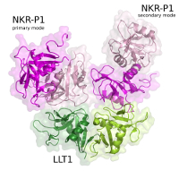
Hsa Siglec and Unique domains in complex with 6S-sialy-Lewisx
Data DOI: 10.15785/SBGRID/787 | Published: 4 Jan 2022
Iverson Laboratory, Vanderbilt University
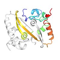
Native dataset for SARS-CoV-2 Nucleocapsid (N) dimerization domain, P21 form
Data DOI: 10.15785/SBGRID/786 | PDB ID 6WZQ: RCSB PDBe | Published: 15 May 2020
Corbett Laboratory, University of California, San Diego
Native dataset for SARS-CoV-2 Nucleocapsid (N) dimerization domain, P1 form
Data DOI: 10.15785/SBGRID/785 | PDB ID 6WZO: RCSB PDBe | Published: 15 May 2020
Corbett Laboratory, University of California, San Diego
Native dataset
Data DOI: 10.15785/SBGRID/782 | PDB ID 6WLY: RCSB PDBe | Published: 19 Jun 2020
Boggon Laboratory, Yale University School of Medicine
Native data
Data DOI: 10.15785/SBGRID/781 | PDB ID 6WLX: RCSB PDBe | Published: 19 Jun 2020
Boggon Laboratory, Yale University School of Medicine
native data set
Data DOI: 10.15785/SBGRID/780 | PDB ID 5MGT: RCSB PDBe | Published: 13 May 2022
Dohnalek Laboratory, Institute of Biotechnology of the Czech Academy of Sciences
native data set
Data DOI: 10.15785/SBGRID/779 | PDB ID 5MGS: RCSB PDBe | Published: 13 May 2022
Dohnalek Laboratory, Institute of Biotechnology of the Czech Academy of Sciences