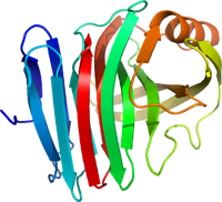
Micro-electron diffraction data from xylanase
Data DOI: 10.15785/SBGRID/286 | PDB ID 5k7p: RCSB PDBe | Published: 31 Mar 2017
Gonen Laboratory, University of California, Los Angeles
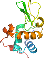
Micro-electron diffraction data from lysozyme
Data DOI: 10.15785/SBGRID/285 | PDB ID 5k7o: RCSB PDBe | Published: 31 Mar 2017
Gonen Laboratory, University of California, Los Angeles
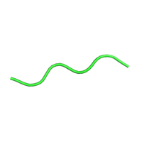
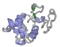
Micro-electron diffraction data from tau VQIVYK peptide
Data DOI: 10.15785/SBGRID/284 | PDB ID 5k7n: RCSB PDBe | Published: 31 Mar 2017
Gonen Laboratory, University of California, Los Angeles
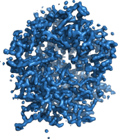
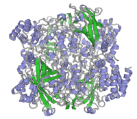
MicroED structure of proteinase K at 1.75 A resolution
Data DOI: 10.15785/SBGRID/262 | PDB ID 5I9S: RCSB PDBe | Published: 31 May 2016
Gonen Laboratory, University of California, Los Angeles
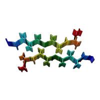
Structure of the amyloid forming segment, GAVVTGVTAVA, from the NAC domain of Parkinson's disease protein alpha-synuclein, residues 68-78, determined by electron diffraction.
Data DOI: 10.15785/SBGRID/223 | PDB ID 4RIL: RCSB PDBe | Publication DOI: 10.1038/nature15368 | Published: 4 Mar 2016
Gonen Laboratory, University of California, Los Angeles
Lysozyme microcrystals
Data DOI: 10.15785/SBGRID/222 | PDB ID 3J6K: RCSB PDBe | Publication DOI: 10.1038/nmeth.3043 | Published: 4 Mar 2016
Gonen Laboratory, University of California, Los Angeles
Bovine catalase microcrystals solved by MicroED
Data DOI: 10.15785/SBGRID/186 | PDB ID 3J7B: RCSB PDBe | Publication DOI: 10.7554/eLife.01345 | Published: 13 Oct 2015
Gonen Laboratory, University of California, Los Angeles