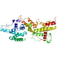
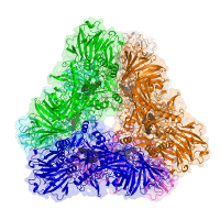
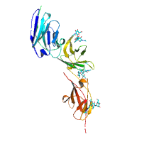
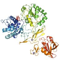
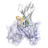
X-Ray Diffraction data from Candida albicans Ras1 guanine-nucleotide exchange factor, source of 7NZZ structure
Data DOI: 10.15785/SBGRID/859 | PDB ID 7NZZ: RCSB PDBe | Published: 25 Jul 2023
Pereira Laboratory, IBMC/i3S, Universidade do Porto
native dataset, P4222 crystal form
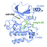
Data DOI: 10.15785/SBGRID/858 | PDB ID 7R85: RCSB PDBe | Published: 9 Nov 2021
Garcia Laboratory, Stanford University
native dataset, P21 crystal form
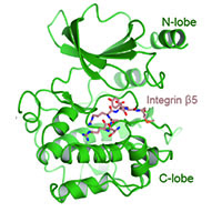
Data DOI: 10.15785/SBGRID/857 | PDB ID 7R84: RCSB PDBe | Published: 9 Nov 2021
Garcia Laboratory, Stanford University
native dataset
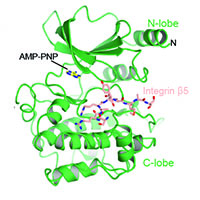
Data DOI: 10.15785/SBGRID/856 | PDB ID 7R86: RCSB PDBe | Published: 9 Nov 2021
Garcia Laboratory, Stanford University
native data set
Data DOI: 10.15785/SBGRID/855 | PDB ID 1YQ2: RCSB PDBe | Publication DOI: 10.1016/j.jmb.2005.08.028 | Published: 29 Oct 2021
Dohnalek Laboratory, Institute of Biotechnology of the Czech Academy of Sciences
Native data
Data DOI: 10.15785/SBGRID/854 | PDB ID 7S47: RCSB PDBe | Published: 18 Nov 2022
Boggon Laboratory, Yale University School of Medicine
Native data
Data DOI: 10.15785/SBGRID/853 | PDB ID 7S48: RCSB PDBe | Published: 18 Nov 2022
Boggon Laboratory, Yale University School of Medicine
Native datasets
Data DOI: 10.15785/SBGRID/852 | PDB ID 7S46: RCSB PDBe | Published: 18 Nov 2022
Boggon Laboratory, Yale University School of Medicine
native dataset
Data DOI: 10.15785/SBGRID/850 | PDB ID 7S2S: RCSB PDBe | Published: 25 Mar 2022
Garcia Laboratory, Stanford University
native dataset
Data DOI: 10.15785/SBGRID/849 | PDB ID 7S2R: RCSB PDBe | Published: 25 Mar 2022
Garcia Laboratory, Stanford University
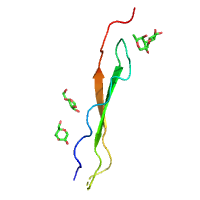
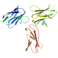
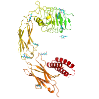
Human thrombin:tsetse thrombin inhibitor complex, source of 7PHX structure
Data DOI: 10.15785/SBGRID/846 | PDB ID 7PHX: RCSB PDBe | Published: 28 Sep 2021
Pereira Laboratory, IBMC/i3S, Universidade do Porto
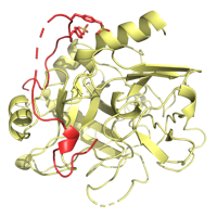
Apo PTP1B by Native S-SAD at Room Temperature
Data DOI: 10.15785/SBGRID/845 | PDB ID 7RIN: RCSB PDBe | Published: 31 Aug 2021
Hekstra Laboratory, Harvard University
Collected at APS on beamline LS-Cat D. 0-360 degrees rotation one degree frames. MARR 300 CCD.
Data DOI: 10.15785/SBGRID/844 | PDB ID 7R7J: RCSB PDBe | Published: 1 Apr 2022
Keck Laboratory, University of Wisconsin-Madison
native dataset
Data DOI: 10.15785/SBGRID/838 | PDB ID 7N3T: RCSB PDBe | Published: 12 Apr 2022
Garcia Laboratory, Stanford University
Native dataset
Data DOI: 10.15785/SBGRID/837 | PDB ID 7R98: RCSB PDBe | Publication DOI: 10.3389/fimmu.2021.719037 | Published: 8 Feb 2022
Corbett Laboratory, University of California, San Diego
Process to 1.42 A
Data DOI: 10.15785/SBGRID/836 | PDB ID 7N0R: RCSB PDBe | Publication DOI: 10.3389/fimmu.2021.719037 | Published: 8 Feb 2022
Corbett Laboratory, University of California, San Diego
Native dataset
Data DOI: 10.15785/SBGRID/835 | PDB ID 7N0I: RCSB PDBe | Publication DOI: 10.3389/fimmu.2021.719037 | Published: 8 Feb 2022
Corbett Laboratory, University of California, San Diego
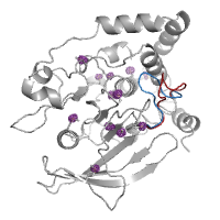
PTP1B in complex with TCS401 by Native S-SAD at Room Temperature
Data DOI: 10.15785/SBGRID/834 | PDB ID 7MM1: RCSB PDBe | Published: 18 May 2021
Hekstra Laboratory, Harvard University
Data collected at the selenium edge of a single crystal of selenomethionine-substituted ClbP grown in a monoolein mesophase.
Data DOI: 10.15785/SBGRID/833 | PDB ID 7MDE: RCSB PDBe | Published: 4 Oct 2022
Gaudet Laboratory, Harvard University
Data collected from a single crystal of the wildtype ClbP peptidase covalently inhibited by a boronic acid and grown in a monopalmitolein mesophase.
Data DOI: 10.15785/SBGRID/832 | PDB ID 7MDC: RCSB PDBe | Published: 4 Oct 2022
Gaudet Laboratory, Harvard University