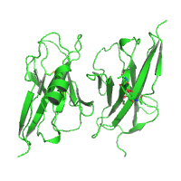
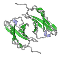
Native dataset
Data DOI: 10.15785/SBGRID/689 | PDB ID 6ONB: RCSB PDBe | Publication DOI: 10.1073/pnas.1818631116 | Published: 6 Dec 2019
Ozkan Laboratory, University of Chicago
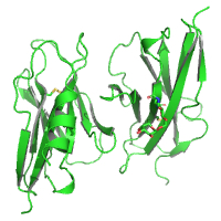
Native dataset
Data DOI: 10.15785/SBGRID/688 | PDB ID 6ON9: RCSB PDBe | Publication DOI: 10.1073/pnas.1818631116 | Published: 6 Dec 2019
Ozkan Laboratory, University of Chicago
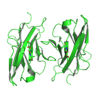
Native dataset
Data DOI: 10.15785/SBGRID/687 | PDB ID 6ON6: RCSB PDBe | Publication DOI: 10.1073/pnas.1818631116 | Published: 6 Dec 2019
Ozkan Laboratory, University of Chicago
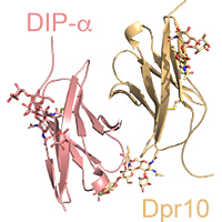
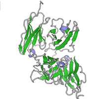
Native dataset
Data DOI: 10.15785/SBGRID/646 | PDB ID 6NRQ: RCSB PDBe | Publication DOI: 10.7554/eLife.41028 | Published: 19 Feb 2019
Ozkan Laboratory, University of Chicago
Native dataset
Data DOI: 10.15785/SBGRID/645 | PDB ID 6NRX: RCSB PDBe | Publication DOI: 10.7554/eLife.41028 | Published: 19 Feb 2019
Ozkan Laboratory, University of Chicago
native dataset
Data DOI: 10.15785/SBGRID/644 | PDB ID 6NS1: RCSB PDBe | Publication DOI: 10.7554/eLife.41028 | Published: 19 Feb 2019
Ozkan Laboratory, University of Chicago
Native dataset
Data DOI: 10.15785/SBGRID/643 | PDB ID 6NRW: RCSB PDBe | Publication DOI: 10.7554/eLife.41028 | Published: 15 Feb 2019
Ozkan Laboratory, University of Chicago
Native dataset
Data DOI: 10.15785/SBGRID/642 | PDB ID 6NRR: RCSB PDBe | Publication DOI: 10.7554/eLife.41028 | Published: 15 Feb 2019
Ozkan Laboratory, University of Chicago
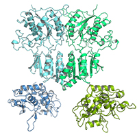
Anisotropic data, with diffraction varying from 3.5 A to 7 A. First 400 images of a 600-image dataset, remainder of which had to be removed due to radiation damage. Crystal is twinned with a twin fraction of 0.49 and twinning law -h, -k, l. True space group is P3(2)21, but appears as P6(x)22 due to twinning.
Data DOI: 10.15785/SBGRID/419 | PDB ID 5L2E: RCSB PDBe | Publication DOI: 10.1016/j.str.2016.11.004 | Published: 20 Jan 2017
Ozkan Laboratory, University of Chicago
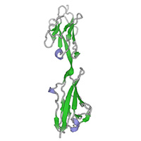
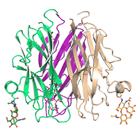
Rat Cerebellin-1, N-terminal domain degraded in the crystallization drops, resulting in the C1q-domain-only trimers.
Data DOI: 10.15785/SBGRID/418 | PDB ID 5KWR: RCSB PDBe | Publication DOI: 10.1016/j.str.2016.11.004 | Published: 17 Jan 2017
Ozkan Laboratory, University of Chicago
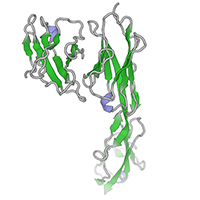
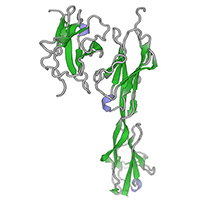
High-resolution native dataset
Data DOI: 10.15785/SBGRID/254 | PDB ID 5EO9: RCSB PDBe | Publication DOI: 10.1016/j.cell.2015.11.022 | Published: 12 Apr 2016
Ozkan Laboratory, University of Chicago