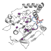
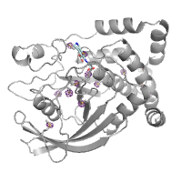
3 passes of 1440 raw diffraction images modeled by PDB ID 9B7C.
Data DOI: 10.15785/SBGRID/1276 | PDB ID 9B7C: RCSB PDBe | Publication DOI: 10.1126/sciadv.adj2921 | Published: 7 Jul 2026
Hekstra Laboratory, Harvard University
A series of 3049 pink-beam, polychromatic still images spaced one degree between adjacent frames. Metadata: Detector distance: 200 mm Beam center: (176.22 mm, 174.68 mm) Peak wavelength: 1.05 Angstroms Bandwidth: 5.5% (BioCARS) Unit cell constants: 79.405, 79.405, 37.837, 90.00, 90.00, 90.00 Space group: P43212
Data DOI: 10.15785/SBGRID/1118 | Published: 9 Jul 2024
Hekstra Laboratory, Harvard University
A series of 180 pink-beam, polychromatic still images spaced one degree between adjacent images. Metadata: Detector distance: 200 mm Beam center: (176.00 mm, 173.93 mm) Peak wavelength: 1.05 Angstroms Beam spectral bandwidth: 5.5% (BioCARS) Unit cell: 34.297, 45.552, 99.035, 90, 90, 90 Space group: P212121
Data DOI: 10.15785/SBGRID/1117 | Published: 9 Jul 2024
Hekstra Laboratory, Harvard University
Time-resolved data consisting of four passes of pink still images for PDZ2. Passes C, D, and E are spaced two degrees between adjacent images, with pass F spaced one degree between adjacent images. Start angles are 0, 92, 181, and 361.5 degrees for passes C, D, E, and F respectively. Time points are collected at 0, 50, 100, and 200 nanoseconds per image per pass. Metadata: Detector distance: 200 mm Beam center: (175.76 mm, 154.17 mm) Peak wavelength: 1.04 Angstroms Beam spectral bandwidth: 5.5% (BioCARS) Unit cell: 65.3, 39.45, 39.01, 90, 117.54, 90 Space group: C121 Applied voltage: 6.01 kV
Data DOI: 10.15785/SBGRID/1116 | PDB ID 5E11: RCSB PDBe | Publication DOI: https://doi.org/10.1038/nature20571 | Published: 9 Jul 2024
Hekstra Laboratory, Harvard University
Apo PTP1B by Native S-SAD at Room Temperature
Data DOI: 10.15785/SBGRID/845 | PDB ID 7RIN: RCSB PDBe | Published: 31 Aug 2021
Hekstra Laboratory, Harvard University
PTP1B in complex with TCS401 by Native S-SAD at Room Temperature
Data DOI: 10.15785/SBGRID/834 | PDB ID 7MM1: RCSB PDBe | Published: 18 May 2021
Hekstra Laboratory, Harvard University
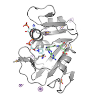
E. coli DHFR by Native Mn,P,S-SAD at Room Temperature
Data DOI: 10.15785/SBGRID/821 | PDB ID 7LVC: RCSB PDBe | Published: 16 Mar 2021
Hekstra Laboratory, Harvard University
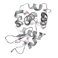
Hen Egg White Lysozyme by Native S-SAD at Room Temperature
Data DOI: 10.15785/SBGRID/816 | PDB ID 7L84: RCSB PDBe | Published: 22 Jan 2021
Hekstra Laboratory, Harvard University