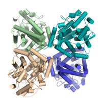
X-Ray Diffraction data from M. hassiacum GgH (Q434F mutant), source of 6G3N structure
Data DOI: 10.15785/SBGRID/1269 | PDB ID 6G3N: RCSB PDBe | Published: 8 May 2026
Pereira Laboratory, IBMC/i3S, Universidade do Porto
Two raw tilt-series (in MRC format) of highly-purified jumbo bacteriophage in association with bacterial flagella. MDOC files are also provided.
Data DOI: 10.15785/SBGRID/1263 | Publication DOI: doi.org/10.1038/s42003-025-09319-7 | Published: 21 Apr 2026
Briegel Laboratory, Institut Pasteur
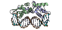
Crystal Structure of the Listeria monocytogenes CadC in complex with DNA
Data DOI: 10.15785/SBGRID/1261 | PDB ID 9T6T: RCSB PDBe | Published: 28 Apr 2026
Cabanes Laboratory, i3S
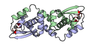
Crystal Structure of the Listeria monocytogenes CadC with Cadmium
Data DOI: 10.15785/SBGRID/1260 | PDB ID 9T6S: RCSB PDBe | Published: 28 Apr 2026
Cabanes Laboratory, i3S
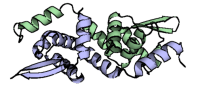
Crystal Structure of the Listeria monocytogenes CadC
Data DOI: 10.15785/SBGRID/1259 | PDB ID 9T6R: RCSB PDBe | Published: 28 Apr 2026
Cabanes Laboratory, i3S
Native dataset. The crystal was macroscopically twinned.
Data DOI: 10.15785/SBGRID/1256 | PDB ID 8EVD: RCSB PDBe | Published: 10 Apr 2026
Madden Laboratory, Dartmouth Geisel School of Medicine
Tilt-series of milled Saccharomyces cerevisiae cells depicting mitochondria, nuclear envelopes, lipid bodies, smooth endoplasmic reticulum and other intracellular components.
Data DOI: 10.15785/SBGRID/1255 | Published: 21 Apr 2026
Raunser Laboratory, Max Planck Institute of Molecular Physiology
Data collected at Vanadium peak wavelength
Data DOI: 10.15785/SBGRID/1235 | Published: 2 Dec 2025
Drennan Laboratory, Massachusetts Institute of Technology
Native dataset
Data DOI: 10.15785/SBGRID/1234 | PDB ID 9PCN: RCSB PDBe | Published: 2 Dec 2025
Drennan Laboratory, Massachusetts Institute of Technology
Native dataset
Data DOI: 10.15785/SBGRID/1233 | PDB ID 9PCM: RCSB PDBe | Published: 2 Dec 2025
Drennan Laboratory, Massachusetts Institute of Technology
Native dataset
Data DOI: 10.15785/SBGRID/1232 | PDB ID 9PCO: RCSB PDBe | Published: 2 Dec 2025
Drennan Laboratory, Massachusetts Institute of Technology
Native dataset
Data DOI: 10.15785/SBGRID/1231 | PDB ID 9PCL: RCSB PDBe | Published: 2 Dec 2025
Drennan Laboratory, Massachusetts Institute of Technology
Native dataset
Data DOI: 10.15785/SBGRID/1230 | PDB ID 9PC5: RCSB PDBe | Published: 2 Dec 2025
Drennan Laboratory, Massachusetts Institute of Technology
Native dataset
Data DOI: 10.15785/SBGRID/1229 | PDB ID 9PBX: RCSB PDBe | Published: 2 Dec 2025
Drennan Laboratory, Massachusetts Institute of Technology
Native dataset
Data DOI: 10.15785/SBGRID/1228 | PDB ID 9PBU: RCSB PDBe | Published: 2 Dec 2025
Drennan Laboratory, Massachusetts Institute of Technology
Native dataset
Data DOI: 10.15785/SBGRID/1227 | PDB ID 9PBN: RCSB PDBe | Published: 2 Dec 2025
Drennan Laboratory, Massachusetts Institute of Technology
Native dataset
Data DOI: 10.15785/SBGRID/1226 | PDB ID 9PBJ: RCSB PDBe | Published: 2 Dec 2025
Drennan Laboratory, Massachusetts Institute of Technology
Native dataset
Data DOI: 10.15785/SBGRID/1225 | PDB ID 9PAY: RCSB PDBe | Published: 2 Dec 2025
Drennan Laboratory, Massachusetts Institute of Technology
Native dataset
Data DOI: 10.15785/SBGRID/1224 | PDB ID 9PAX: RCSB PDBe | Published: 2 Dec 2025
Drennan Laboratory, Massachusetts Institute of Technology
Native dataset
Data DOI: 10.15785/SBGRID/1223 | PDB ID 9PAU: RCSB PDBe | Published: 2 Dec 2025
Drennan Laboratory, Massachusetts Institute of Technology