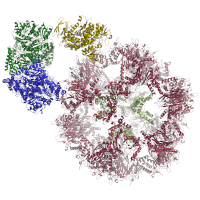
Structural model of the E3BP core, refined in a C2 symmetrized cryoEM map. E3BP showed a minimal fold, which is conserved, and can be found in various other E2 proteins from diverse acyl-transferases, including the 2-keto acid dehydrogenase family. Additionally, by fitting the structural models into an asymmetrically refined cryoEM map, we supply a structural model for the native core scaffold of the PDHc metabolon from C. thermophilum. Models were built and refined by COOT and PHENIX. To capture the transient interaction of lipoyl domain (LD) and the core structure during the transacetylase reaction, we docked the LDs of C. thermophilum, H. sapiens, and N. crassa to their respective core structure. HADDOCK parameter files are deposited to reproduce docking. The best docking solution of C. thermophilum was used to study the interaction by extensive MD simulations. Parameter files and results are also given, to reproduce these simulations.
Data DOI: 10.15785/SBGRID/848 | PDB ID 7OTT: RCSB PDBe | Published: 23 Nov 2021
Kastritis Laboratory, Martin Luther University Halle-Wittenberg
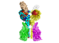
Structural models of the E1 and E2 proteins of Pyruvate Dehydrogenase Complex (PDHc), 2-Oxoglutarate Dehydrogenase (OGDHc), and Branched-Chain alpha-Keto Acid Dehydrogenase Complex (BCKDHc), E3BP of PDHc and E3, shared among all three complexes. In addition, a cif-file of E1, E2, E3BP, and E3 of PDHc modeled from cryoEM data is provided. Models were generated by homology modeling using MODELLER and refined using HADDOCK webserver.
Data DOI: 10.15785/SBGRID/817 | PDB ID 7BGJ: RCSB PDBe | Published: 26 Jan 2021
Kastritis Laboratory, Martin Luther University Halle-Wittenberg
242 structural models of human ACE2 variants with the bound RBD of SARS-CoV-2 Spike glycoprotein refined with HADDOCK2.2 (initial PDB: 6M17, 6M0J). The structural models include in human population naturally occurring variants of ACE2 (140), models of the mutants with reported effects on the recognition of RBD (39), and computational alanine scanning mutagenesis of ACE2-RBD interface (63). Moreover, all aforementioned ACE2 variants can be found as a structural models of ACE2-B(0)AT1 complex with the bound RBD, including all ions and structurally-important glycan molecules (initial PDB: 6M17).
Data DOI: 10.15785/SBGRID/791 | Published: 21 Aug 2020
Kastritis Laboratory, Martin Luther University Halle-Wittenberg