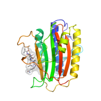
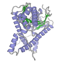
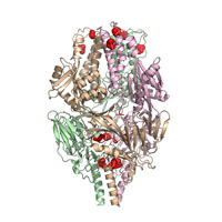
Native data for C. elegans HTP-1 bound to HTP-3 motif-1
Data DOI: 10.15785/SBGRID/209 | PDB ID 4TZQ: RCSB PDBe | Publication DOI: 10.1016/j.devcel.2014.09.013 | Published: 18 Dec 2015
Corbett Laboratory, University of California, San Diego
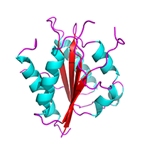
Native data for C. elegans HTP-1 bound to HIM-3 closure motif
Data DOI: 10.15785/SBGRID/208 | PDB ID 4TZO: RCSB PDBe | Publication DOI: 10.1016/j.devcel.2014.09.013 | Published: 18 Dec 2015
Corbett Laboratory, University of California, San Diego
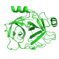
Native data for C. elegans HTP-2 bound to HTP-3 motif-6
Data DOI: 10.15785/SBGRID/207 | PDB ID 4TZN: RCSB PDBe | Publication DOI: 10.1016/j.devcel.2014.09.013 | Published: 18 Dec 2015
Corbett Laboratory, University of California, San Diego
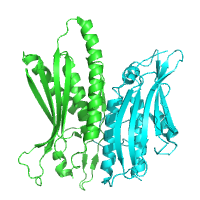
Native data for C. elegans HTP-2 bound to HTP-3 closure motif 1
Data DOI: 10.15785/SBGRID/206 | PDB ID 4TZM: RCSB PDBe | Publication DOI: 10.1016/j.devcel.2014.09.013 | Published: 18 Dec 2015
Corbett Laboratory, University of California, San Diego
Native data for C. elegans HTP-2 bound to HIM-3 closure motif, P21 form
Data DOI: 10.15785/SBGRID/205 | PDB ID 4TZL: RCSB PDBe | Publication DOI: 10.1016/j.devcel.2014.09.013 | Published: 18 Dec 2015
Corbett Laboratory, University of California, San Diego
Native data for C. elegans HIM-3 bound to HTP-3 closure motif-4
Data DOI: 10.15785/SBGRID/204 | PDB ID 4TZJ: RCSB PDBe | Publication DOI: 10.1016/j.devcel.2014.09.013 | Published: 18 Dec 2015
Corbett Laboratory, University of California, San Diego
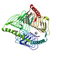
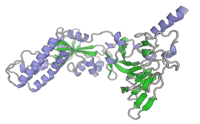
Native diffraction dataset of B. abortus WrpA protein
Data DOI: 10.15785/SBGRID/203 | PDB ID 5F51: RCSB PDBe | Publication DOI: 10.1128/JB.00982-15 | Published: 15 Dec 2015
Crosson Laboratory, University of Chicago
SeMet dataset
Data DOI: 10.15785/SBGRID/202 | PDB ID 5F4B: RCSB PDBe | Publication DOI: 10.1128/JB.00982-15 | Published: 15 Dec 2015
Crosson Laboratory, University of Chicago
Trypsin crystals were obtained according to the method of Liebschner et.al (Liebschner et al., 2013). Lyophilized bovine pancreas trypsin was purchased from Sigma-Aldrich (T1005) and dissolved at a concentration of 30 mg/mL into 30mM HEPES pH 7.0, 5 mg/mL benzamidine and 3mM CaCl2. Crystals were obtained from a solution of 200mM Ammonium sulfate, 100mM Na cacodylate pH 6.5, 20% PEG 8000 and 15% glycerol. Data collected for simultaneous Bragg and Diffuse Scattering processing.
Data DOI: 10.15785/SBGRID/201 | PDB ID 5F6M: RCSB PDBe | Published: 18 Dec 2015
Fraser Laboratory, University of California, San Francisco
Native dataset
Data DOI: 10.15785/SBGRID/200 | PDB ID 5C50: RCSB PDBe | Publication DOI: 10.1016/j.str.2015.07.011 | Published: 4 Dec 2015
Hurley Laboratory, University of California, Berkeley
NaBr soaked dataset
Data DOI: 10.15785/SBGRID/199 | PDB ID 5C50: RCSB PDBe | Publication DOI: 10.1016/j.str.2015.07.011 | Published: 4 Dec 2015
Hurley Laboratory, University of California, Berkeley
Iodine SAD dataset collected at 12670 electron volts. Crystal diffraction was measured at a temperature of 100 K using a 1 degree oscillation range.
Data DOI: 10.15785/SBGRID/198 | PDB ID 5CEG: RCSB PDBe | Publication DOI: 10.1016/j.cell.2015.09.055 | Published: 24 Nov 2015
Crosson Laboratory, University of Chicago
Diffraction data from this crystal of B. abortus RicA were collected at a temperature of 100 K using a 1° oscillation range.
Data DOI: 10.15785/SBGRID/197 | PDB ID 4N27: RCSB PDBe | Publication DOI: 10.1021/bi401373r | Published: 24 Nov 2015
Crosson Laboratory, University of Chicago
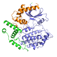
Native dataset for Hrr25:Mam1 complex, form 2
Data DOI: 10.15785/SBGRID/196 | PDB ID 5CZO: RCSB PDBe | Published: 9 Aug 2016
Corbett Laboratory, University of California, San Diego
Native diffraction data for S. cerevisiae Hrr25:Mam1, form 1
Data DOI: 10.15785/SBGRID/195 | PDB ID 5CYZ: RCSB PDBe | Published: 9 Aug 2016
Corbett Laboratory, University of California, San Diego
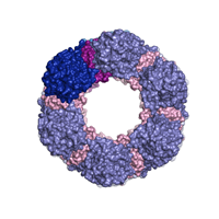
Native dataset
Data DOI: 10.15785/SBGRID/194 | PDB ID 5CXF: RCSB PDBe | Publication DOI: 10.1371/journal.ppat.1005227 | Published: 13 Nov 2015
Heldwein Laboratory, Tufts University School of Medicine
Native dataset
Data DOI: 10.15785/SBGRID/192 | PDB ID 5C6B: RCSB PDBe | Publication DOI: 10.1038/ncomms9143 | Published: 30 Oct 2015
McLellan Laboratory, University of Texas at Austin
Native dataset
Data DOI: 10.15785/SBGRID/191 | PDB ID 5C69: RCSB PDBe | Publication DOI: 10.1038/ncomms9143 | Published: 30 Oct 2015
McLellan Laboratory, University of Texas at Austin
Native dataset.
Data DOI: 10.15785/SBGRID/189 | PDB ID 4ZXS: RCSB PDBe | Publication DOI: 10.15252/embj.201592359 | Published: 30 Oct 2015
Heldwein Laboratory, Tufts University School of Medicine
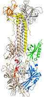
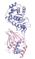
Soluble portion of pseudorabies virus nuclear egress complex.
Data DOI: 10.15785/SBGRID/188 | PDB ID 4Z3U: RCSB PDBe | Publication DOI: 10.15252/embj.201592359 | Published: 30 Oct 2015
Heldwein Laboratory, Tufts University School of Medicine